
Search Count: 63
 |
Organism: Juglans regia
Method: X-RAY DIFFRACTION Resolution:3.10 Å Release Date: 2023-12-20 Classification: PROTEIN BINDING |
 |
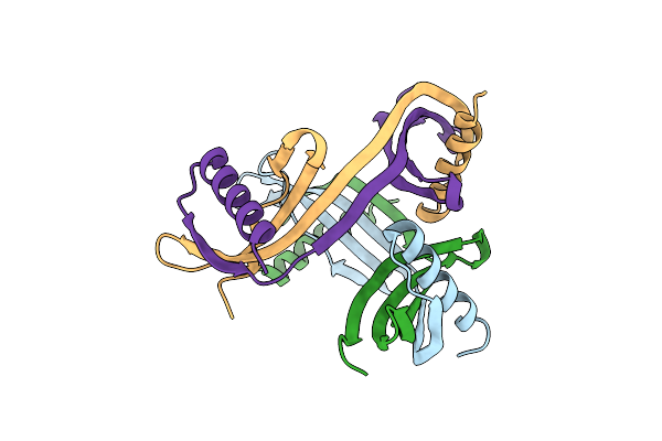
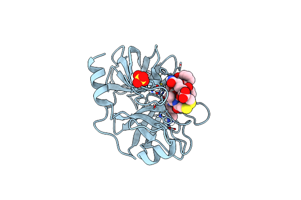
Structure Of Topi1 Inhibitor From Tityus Obscurus Scorpion Venom In Complex With Trypsin
Organism: Bos taurus, Tityus
Method: X-RAY DIFFRACTION Resolution:1.29 Å Release Date: 2020-07-01 Classification: hydrolase/hydrolase inhibitor Ligands: CA, SO4 |
 |
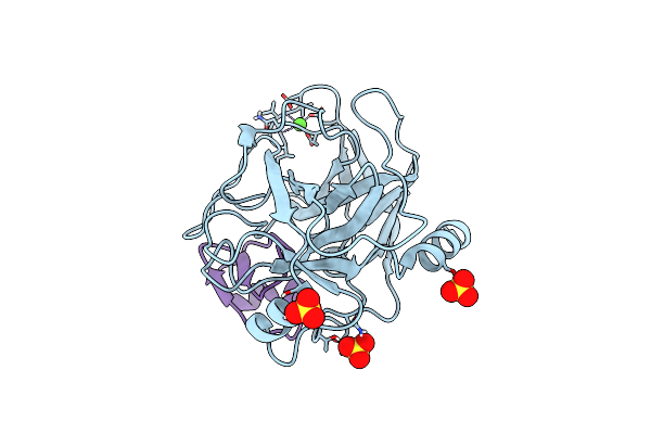
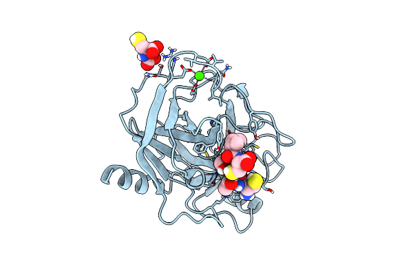
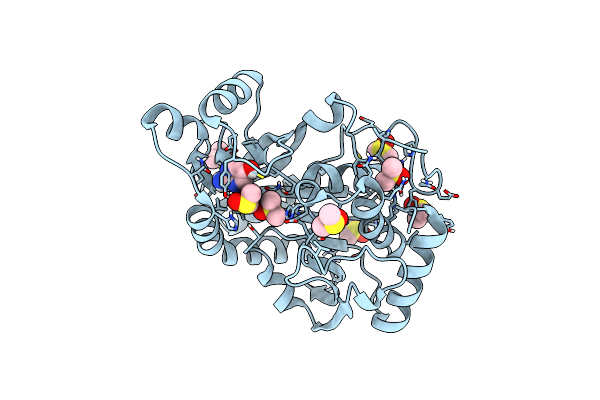
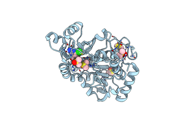
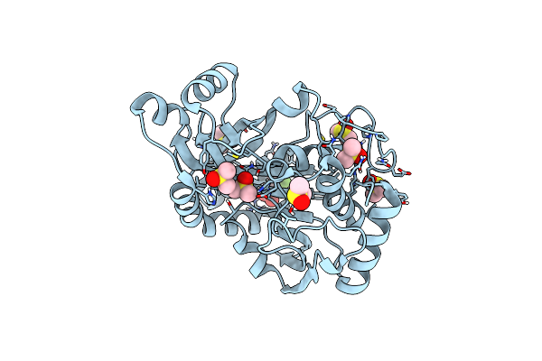
Crystallographic Structure Of The Octapeptide Derived From The Btci Inhibitor Bound To Beta-Trypsin In Space Group P 21 21 21.
Organism: Vigna unguiculata, Bos taurus
Method: X-RAY DIFFRACTION Resolution:1.18 Å Release Date: 2019-08-07 Classification: hydrolase/hydrolase inhibitor Ligands: SO4, CA |
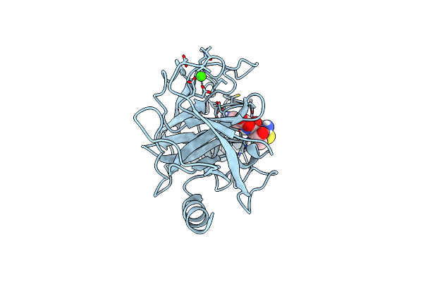 |
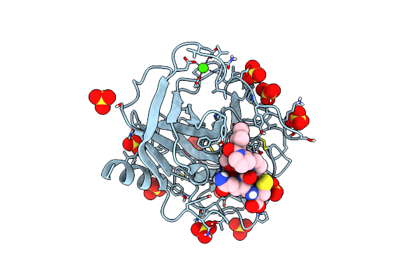
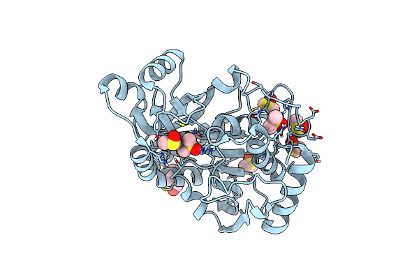
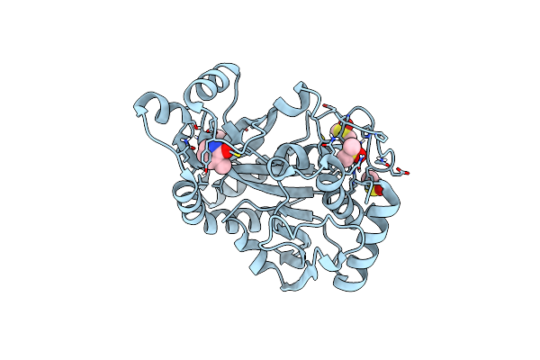
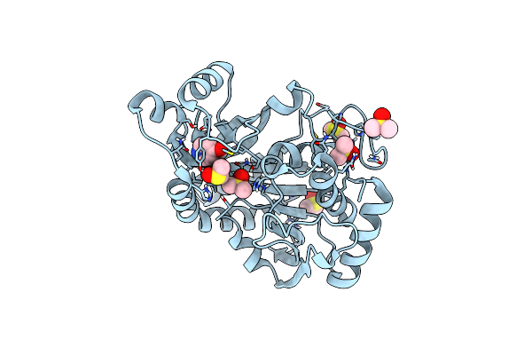
Crystallographic Structure Of The Cyclic Heptapeptide Derived From The Btci Inhibitor Bound To Beta-Trypsin In Space Group P 4(1) 2(1) 2
Organism: Vigna unguiculata, Bos taurus
Method: X-RAY DIFFRACTION Resolution:1.39 Å Release Date: 2019-08-07 Classification: hydrolase/hydrolase inhibitor Ligands: CA |
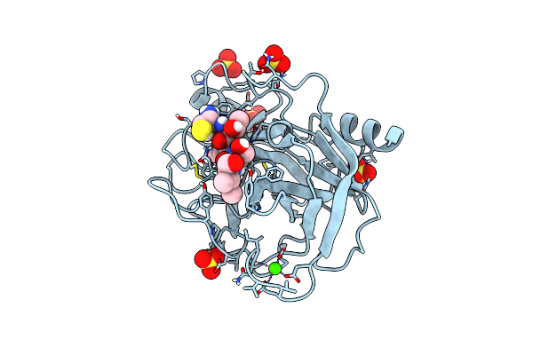 |
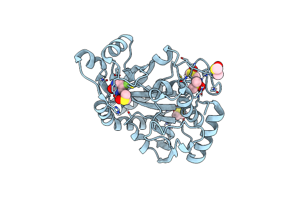
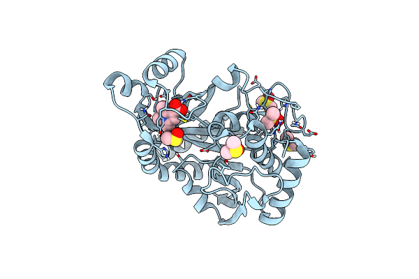
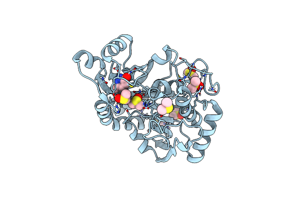
Crystallographic Structure Of The Cyclic Heptapeptide Derived From The Btci Inhibitor Bound To Beta-Trypsin In Space Group P 21 21 21
Organism: Vigna unguiculata, Bos taurus
Method: X-RAY DIFFRACTION Resolution:1.29 Å Release Date: 2019-08-07 Classification: hydrolase/hydrolase inhibitor Ligands: SO4, CA |
 |
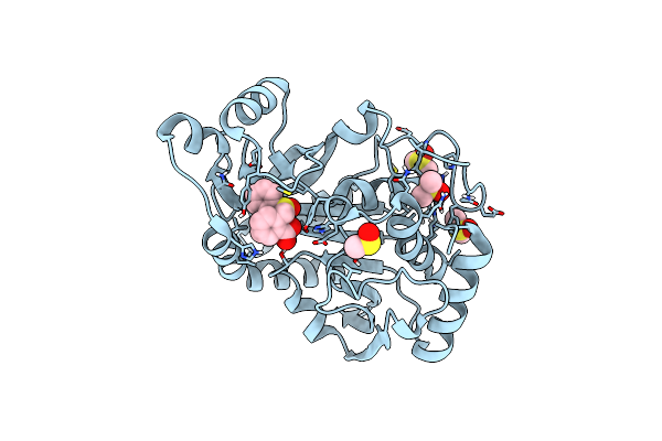
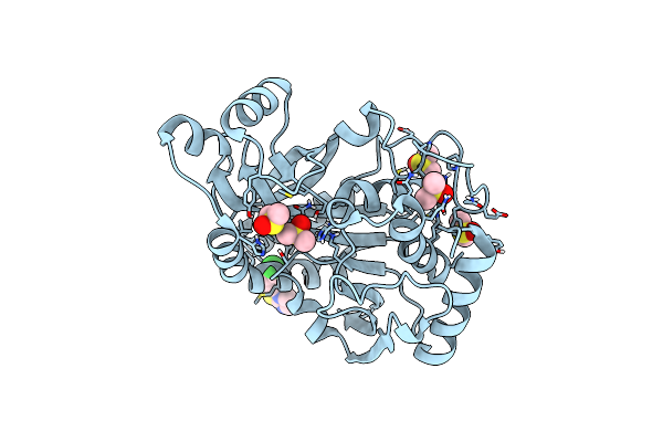
Crystallographic Structure Of The Cyclic Hexapeptide Derived From The Btci Inhibitor Bound To Beta-Trypsin In Space Group P 21 21 21
Organism: Vigna unguiculata, Bos taurus
Method: X-RAY DIFFRACTION Resolution:1.19 Å Release Date: 2019-08-07 Classification: hydrolase/hydrolase inhibitor Ligands: CA, J3D |
 |
Crystallographic Structure Of The Cyclic Nonapeptide Derived From The Btci Inhibitor Bound To Beta-Trypsin In Space Group P 32 2 1
Organism: Vigna unguiculata, Bos taurus
Method: X-RAY DIFFRACTION Resolution:1.61 Å Release Date: 2019-03-13 Classification: hydrolase/hydrolase inhibitor Ligands: CA, SO4 |
 |
Crystallographic Structure Of The Cyclic Nonapeptide Derived From The Btci Inhibitor Bound To Beta-Trypsin In Space Group P 21 21 21.
Organism: Vigna unguiculata, Bos taurus
Method: X-RAY DIFFRACTION Resolution:1.15 Å Release Date: 2019-03-13 Classification: hydrolase/hydrolase inhibitor Ligands: CA, SO4 |
 |
Crystal Structure Of Purine Nucleoside Phosphorylase Isoform 2 From Schistosoma Mansoni In Complex With 5-Fluoro-1,2-Dihydropyrimidin-2-One
Organism: Schistosoma mansoni
Method: X-RAY DIFFRACTION Resolution:1.92 Å Release Date: 2018-11-07 Classification: TRANSFERASE Ligands: DMS, DJP |
 |
Crystal Structure Of Purine Nucleoside Phosphorylase Isoform 2 From Schistosoma Mansoni In Complex With 2-Aminopyrimidin-5-Ol
Organism: Schistosoma mansoni
Method: X-RAY DIFFRACTION Resolution:2.00 Å Release Date: 2018-11-07 Classification: TRANSFERASE Ligands: DMS, DS1 |
 |
Crystal Structure Of Purine Nucleoside Phosphorylase Isoform 2 From Schistosoma Mansoni In Complex With (2R)-2-Amino-3-(Benzyloxy)Propan-1-Ol
Organism: Schistosoma mansoni
Method: X-RAY DIFFRACTION Resolution:1.42 Å Release Date: 2018-11-07 Classification: TRANSFERASE Ligands: DMS, SEM |
 |
Crystal Structure Of Purine Nucleoside Phosphorylase Isoform 2 From Schistosoma Mansoni In Complex With 1,2,5-Trimethyl-1H-Pyrrole-3-Carboxylic Acid
Organism: Schistosoma mansoni
Method: X-RAY DIFFRACTION Resolution:1.59 Å Release Date: 2018-11-07 Classification: TRANSFERASE Ligands: DMS, DS7 |
 |
Crystal Structure Of Purine Nucleoside Phosphorylase Isoform 2 From Schistosoma Mansoni In Complex With 1-(4-Amino-2-Hydroxyphenyl)Ethan-1-One
Organism: Schistosoma mansoni
Method: X-RAY DIFFRACTION Resolution:1.60 Å Release Date: 2018-11-07 Classification: TRANSFERASE Ligands: DMS, DSJ |
 |
Crystal Structure Of Purine Nucleoside Phosphorylase Isoform 2 From Schistosoma Mansoni In Complex With 2-{[(S)-Benzenesulfinyl]Methyl}Benzoic Acid
Organism: Schistosoma mansoni
Method: X-RAY DIFFRACTION Resolution:1.73 Å Release Date: 2018-11-07 Classification: TRANSFERASE Ligands: DMS, DTS |
 |
Crystal Structure Of Purine Nucleoside Phosphorylase Isoform 2 From Schistosoma Mansoni In Complex With 4-Chloro-6-Methylpyrimidin-2-Amine
Organism: Schistosoma mansoni
Method: X-RAY DIFFRACTION Resolution:1.56 Å Release Date: 2018-11-07 Classification: TRANSFERASE Ligands: DMS, DU7 |
 |
Crystal Structure Of Purine Nucleoside Phosphorylase Isoform 2 From Schistosoma Mansoni In Complex With 3-Methyl-1,2-Dihydropyridin-2-One
Organism: Schistosoma mansoni
Method: X-RAY DIFFRACTION Resolution:1.44 Å Release Date: 2018-10-17 Classification: TRANSFERASE/INHIBITOR Ligands: DMS, D4V |
 |
Crystal Structure Of Purine Nucleoside Phosphorylase Isoform 2 From Schistosoma Mansoni In Complex With 6-Methyl-2,3-Dihydropyridazin-3-One
Organism: Schistosoma mansoni
Method: X-RAY DIFFRACTION Resolution:1.46 Å Release Date: 2018-10-10 Classification: TRANSFERASE/INHIBITOR Ligands: DMS, CVG |
 |
Crystal Structure Of Purine Nucleoside Phosphorylase Isoform 2 From Schistosoma Mansoni In Complex With3-(4-Chlorophenyl)-5H,6H-Imidazo[2,1-B][1,3]Thiazole
Organism: Schistosoma mansoni
Method: X-RAY DIFFRACTION Resolution:1.52 Å Release Date: 2018-10-10 Classification: TRANSFERASE/INHIBITOR Ligands: DMS, A5H |
 |
Crystal Structure Of Purine Nucleoside Phosphorylase Isoform 2 From Schistosoma Mansoni In Complex With (2S)-2-(3,5-Difluorophenyl)-2-Hydroxyacetic Acid
Organism: Schistosoma mansoni
Method: X-RAY DIFFRACTION Resolution:1.54 Å Release Date: 2018-10-10 Classification: TRANSFERASE/INHIBITOR Ligands: DMS, CXJ |
 |
Crystal Structure Of Purine Nucleoside Phosphorylase Isoform 2 From Schistosoma Mansoni In Complex With Pyridin-4-Ol
Organism: Schistosoma mansoni
Method: X-RAY DIFFRACTION Resolution:1.60 Å Release Date: 2018-10-03 Classification: TRANSFERASE/INHIBITOR Ligands: DMS, CQG |