
Search Count: 30
 |
Organism: Claviceps fusiformis
Method: ELECTRON MICROSCOPY Release Date: 2025-01-01 Classification: OXIDOREDUCTASE Ligands: HEM |
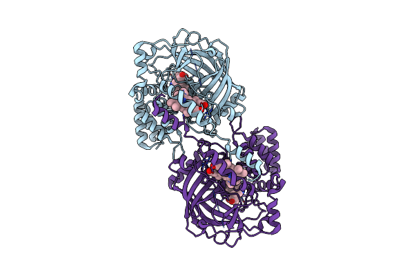 |
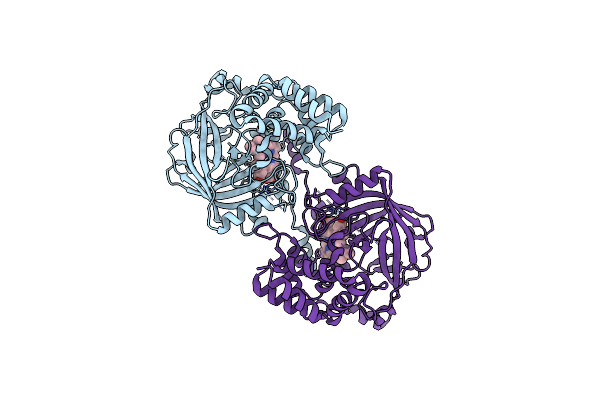
Structure Of Chanoclavine Synthase From Claviceps Fusiformis In Complex With Prechanoclavine
Organism: Claviceps fusiformis
Method: ELECTRON MICROSCOPY Resolution:2.33 Å Release Date: 2025-01-01 Classification: OXIDOREDUCTASE Ligands: HEM, LH6 |
 |
Organism: Homo sapiens
Method: X-RAY DIFFRACTION Resolution:1.40 Å Release Date: 2022-08-24 Classification: TRANSFERASE/TRANSFERASE INHIBITOR Ligands: B4I |
 |
Organism: Severe acute respiratory syndrome coronavirus 2, Vicugna pacos
Method: X-RAY DIFFRACTION Resolution:3.00 Å Release Date: 2022-06-29 Classification: VIRAL PROTEIN Ligands: GOL, PO4 |
 |
Organism: Severe acute respiratory syndrome coronavirus 2, Vicugna pacos
Method: X-RAY DIFFRACTION Resolution:1.75 Å Release Date: 2022-05-25 Classification: VIRAL PROTEIN Ligands: GOL, ACT |
 |
Cryo-Em Structure Of The Human Glycosylphosphatidylinositol Transamidase Complex At 2.53 Angstrom Resolution
Organism: Homo sapiens
Method: ELECTRON MICROSCOPY Resolution:2.53 Å Release Date: 2022-04-27 Classification: TRANSFERASE Ligands: Y01, AJP, BJR, PEE, CA, P5S, NAG, CLR, 05E, PA1, CDL, DKB, 06O |
 |
Cryo-Em Structure Of The Hedgehog Release Protein Disp From Water Bear (Hypsibius Dujardini)
Organism: Hypsibius dujardini
Method: ELECTRON MICROSCOPY Release Date: 2021-09-08 Classification: MEMBRANE PROTEIN |
 |
Structure Of Sybody Sr4 In Complex With The Sars-Cov-2 S Receptor Binding Domain (Rbd)
Organism: Synthetic construct, Severe acute respiratory syndrome coronavirus 2
Method: X-RAY DIFFRACTION Resolution:2.15 Å Release Date: 2020-06-24 Classification: PROTEIN BINDING Ligands: GOL, NAG |
 |
Structure Of Sybody Mr17 In Complex With The Sars-Cov-2 S Receptor-Binding Domain (Rbd)
Organism: Synthetic construct, Severe acute respiratory syndrome coronavirus 2
Method: X-RAY DIFFRACTION Resolution:2.77 Å Release Date: 2020-06-24 Classification: PROTEIN BINDING Ligands: GOL |
 |
Structure Of Sybody Mr17-K99Y In Complex With The Sars-Cov-2 S Receptor-Binding Domain (Rbd)
Organism: Synthetic construct, Severe acute respiratory syndrome coronavirus 2
Method: X-RAY DIFFRACTION Resolution:2.94 Å Release Date: 2020-06-24 Classification: PROTEIN BINDING Ligands: GOL |
 |
Crystal Structure Of Plsy (Ygih), An Integral Membrane Glycerol 3-Phosphate Acyltransferase - The Monoacylglycerol Form
Organism: Aquifex aeolicus
Method: X-RAY DIFFRACTION Resolution:1.48 Å Release Date: 2017-12-06 Classification: TRANSFERASE Ligands: SO4, 78M, PGE, GLY |
 |
Crystal Structure Of Plsy (Ygih), An Integral Membrane Glycerol 3-Phosphate Acyltransferase - The Glycerol 3-Phosphate Form
Organism: Aquifex aeolicus
Method: X-RAY DIFFRACTION Resolution:2.37 Å Release Date: 2017-12-06 Classification: TRANSFERASE Ligands: G3P, OLC, PO4 |
 |
Crystal Structure Of Plsy (Ygih), An Integral Membrane Glycerol 3-Phosphate Acyltransferase - The Acyl Phosphate Form
Organism: Aquifex aeolicus
Method: X-RAY DIFFRACTION Resolution:1.77 Å Release Date: 2017-12-06 Classification: TRANSFERASE Ligands: 78M, 87O, K, PO4 |
 |
Crystal Structure Of Plsy (Ygih), An Integral Membrane Glycerol 3-Phosphate Acyltransferase - The Lysphosphatidic Acid Form
Organism: Aquifex aeolicus
Method: X-RAY DIFFRACTION Resolution:2.41 Å Release Date: 2017-12-06 Classification: TRANSFERASE Ligands: PO4, NKO |
 |
Crystal Structure Of Plsy (Ygih), An Integral Membrane Glycerol 3-Phosphate Acyltransferase - The Orthophosphate Form
Organism: Aquifex aeolicus
Method: X-RAY DIFFRACTION Resolution:1.83 Å Release Date: 2017-12-06 Classification: TRANSFERASE Ligands: PO4, 78M |
 |
Organism: Homo sapiens
Method: SOLUTION NMR Release Date: 2014-10-29 Classification: METAL BINDING PROTEIN Ligands: ZN |
 |
Organism: Homo sapiens
Method: X-RAY DIFFRACTION Resolution:1.76 Å Release Date: 2014-10-29 Classification: LIGASE Ligands: ZN |
 |
Organism: Drosophila melanogaster
Method: SOLUTION NMR Release Date: 2014-03-05 Classification: ONCOPROTEIN |
 |
Organism: Human betacoronavirus 2c emc/2012
Method: X-RAY DIFFRACTION Resolution:2.51 Å Release Date: 2013-07-10 Classification: VIRAL PROTEIN Ligands: NAG |
 |
Organism: Homo sapiens, Human betacoronavirus 2c emc/2012
Method: X-RAY DIFFRACTION Resolution:2.70 Å Release Date: 2013-07-10 Classification: HYDROLASE/VIRAL PROTEIN Ligands: NAG |