
Search Count: 20
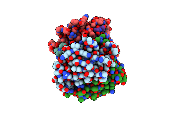 |
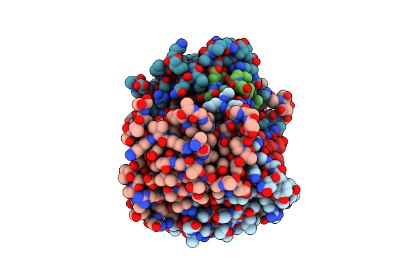
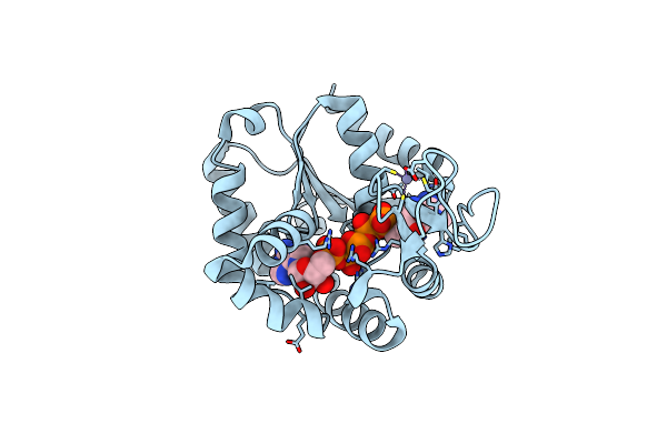
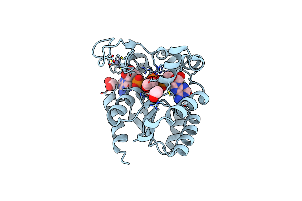
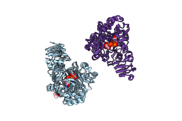
Crystal Structure Of Streptavidin With Peptide D-Amino Acid Containing Peptide Gyglanvdessg
Organism: Streptomyces avidinii, Synthetic construct
Method: X-RAY DIFFRACTION Resolution:1.05 Å Release Date: 2017-10-25 Classification: STREPTAVIDIN PEPTIDE 11101 COMPLEX Ligands: IPA |
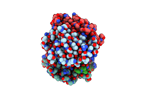 |
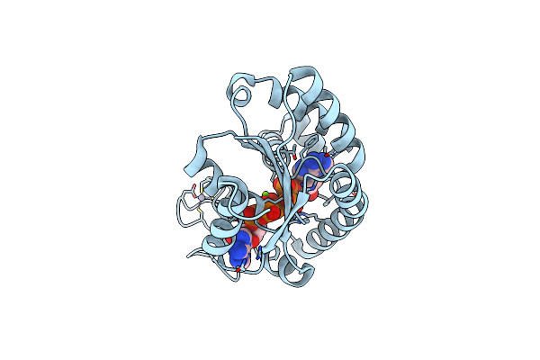
Organism: Streptomyces avidinii, Synthetic construct
Method: X-RAY DIFFRACTION Resolution:1.10 Å Release Date: 2017-10-11 Classification: BIOTIN BINDING PROTEIN |
 |
Organism: Streptomyces avidinii, Homo sapiens
Method: X-RAY DIFFRACTION Resolution:1.12 Å Release Date: 2017-10-04 Classification: BIOTIN BINDING PROTEIN |
 |
Organism: Streptomyces avidinii, Synthetic construct
Method: X-RAY DIFFRACTION Resolution:1.27 Å Release Date: 2017-10-04 Classification: biotin binding protein Ligands: GOL |
 |
Organism: Streptomyces avidinii, Synthetic construct
Method: X-RAY DIFFRACTION Resolution:1.03 Å Release Date: 2017-10-04 Classification: BIOTIN BINDING PROTEIN |
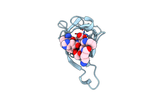 |
Crystal Structure Of Streptavidin D-Amino Acid Containing Peptide Gdlwqheatwkkq
Organism: Streptomyces avidinii
Method: X-RAY DIFFRACTION Resolution:1.61 Å Release Date: 2017-10-04 Classification: Biotin binding protein Ligands: DLE, DTR, DGN, DHI, DGL, DAL, DTH, DLY |
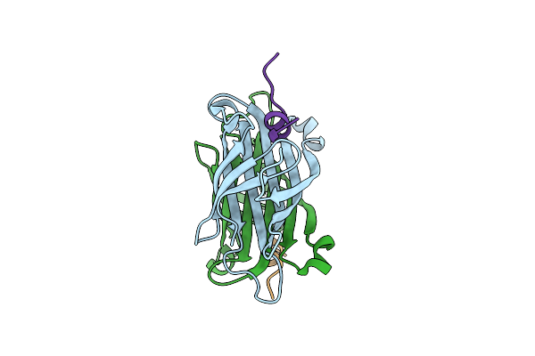 |
Crystal Structure Of Streptavidin With D-Amino Acid Containing Peptide Ggwhdeatwkpg
Organism: Streptomyces avidinii, Synthetic construct
Method: X-RAY DIFFRACTION Resolution:1.10 Å Release Date: 2017-10-04 Classification: biotin binding protein |
 |
Organism: Streptomyces avidinii, Synthetic construct
Method: X-RAY DIFFRACTION Resolution:1.50 Å Release Date: 2017-10-04 Classification: Biotin binding protein |
 |
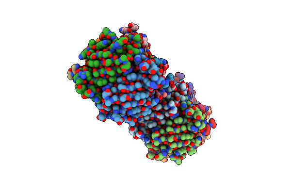
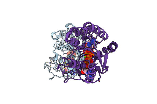
Organism: Bacillus subtilis
Method: X-RAY DIFFRACTION Resolution:1.80 Å Release Date: 2014-06-25 Classification: TRANSFERASE Ligands: ZN, AP5 |
 |
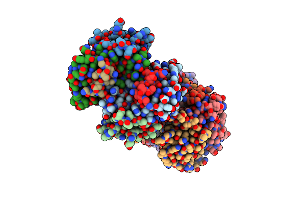
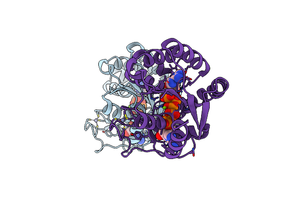
Organism: Bacillus subtilis
Method: X-RAY DIFFRACTION Resolution:1.37 Å Release Date: 2014-06-25 Classification: TRANSFERASE Ligands: ZN, AP5, MG |
 |
Organism: Geobacillus stearothermophilus
Method: X-RAY DIFFRACTION Resolution:1.67 Å Release Date: 2014-06-25 Classification: TRANSFERASE Ligands: AP5, ZN, MG |
 |
Organism: Geobacillus stearothermophilus
Method: X-RAY DIFFRACTION Resolution:1.67 Å Release Date: 2014-06-25 Classification: TRANSFERASE Ligands: ZN, MG, AP5 |
 |
Organism: Bacillus subtilis
Method: X-RAY DIFFRACTION Resolution:1.82 Å Release Date: 2009-06-09 Classification: TRANSFERASE Ligands: ZN, MG, AP5, EDO |
 |
Organism: Bacillus subtilis
Method: X-RAY DIFFRACTION Resolution:1.58 Å Release Date: 2009-06-09 Classification: TRANSFERASE Ligands: ZN, MG, AP5 |
 |
Organism: Homo sapiens
Method: X-RAY DIFFRACTION Resolution:3.00 Å Release Date: 2008-01-15 Classification: CHAPERONE Ligands: ZN, SO4 |
 |
Organism: Danio rerio
Method: X-RAY DIFFRACTION Resolution:2.60 Å Release Date: 2006-10-10 Classification: ENDOCYTOSIS/EXOCYTOSIS Ligands: SO4 |
 |
Crystal Structure Of A Putative Udp-Glucose Pyrophosphorylase From Arabidopsis Thaliana With Bound Udp-Glucose
Organism: Arabidopsis thaliana
Method: X-RAY DIFFRACTION Resolution:1.64 Å Release Date: 2006-10-03 Classification: TRANSFERASE Ligands: DMS, UPG |
 |
Crystal Structure Of A Putative Udp-Glucose Pyrophosphorylase From Arabidopsis Thaliana With Bound Utp
Organism: Arabidopsis thaliana
Method: X-RAY DIFFRACTION Resolution:1.85 Å Release Date: 2006-09-26 Classification: TRANSFERASE Ligands: DMS, UTP |
 |
X-Ray Structure Of Adenosine 5'-Monophosphate Deaminase From Arabidopsis Thaliana In Complex With Coformycin 5'-Phosphate
Organism: Arabidopsis thaliana
Method: X-RAY DIFFRACTION Resolution:3.34 Å Release Date: 2005-07-19 Classification: HYDROLASE Ligands: ZN, PO4, CF5 |
 |
X-Ray Structure Of Gene Product From Arabidopsis Thaliana At3G03250, A Putative Udp-Glucose Pyrophosphorylase
Organism: Arabidopsis thaliana
Method: X-RAY DIFFRACTION Resolution:1.86 Å Release Date: 2005-04-12 Classification: STRUCTURAL GENOMICS, UNKNOWN FUNCTION |