
Search Count: 26
 |
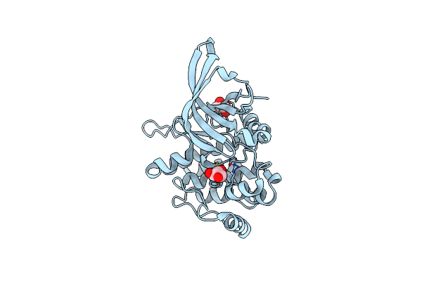
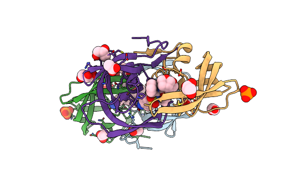
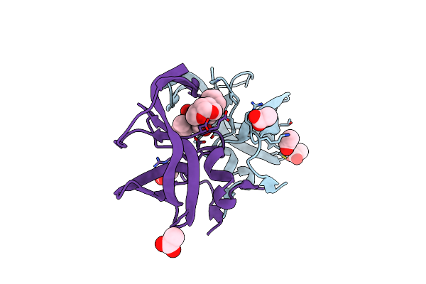
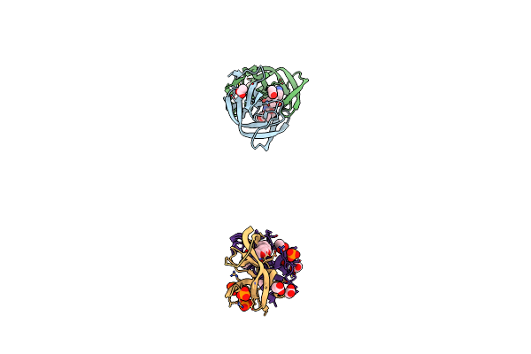
Mutations Outside The Active Site Of Hiv-1 Protease Alter Enzyme Structure And Dynamic Ensemble Of The Active Site To Confer Drug Resistance
Organism: Human immunodeficiency virus 1
Method: X-RAY DIFFRACTION Resolution:1.85 Å Release Date: 2015-02-18 Classification: hydrolase/hydrolase inhibitor Ligands: PO4, 017, ACT |
 |
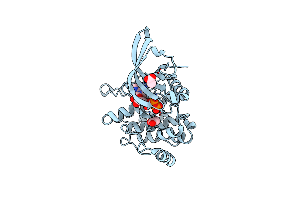
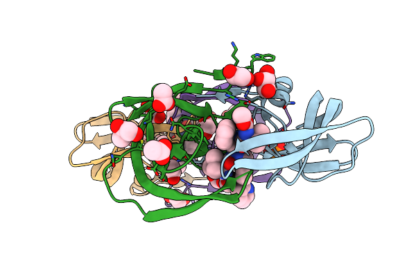
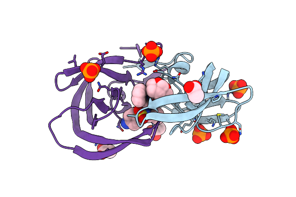
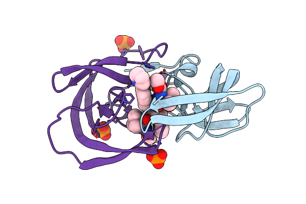
Mutations Outside The Active Site Of Hiv-1 Protease Alter Enzyme Structure And Dynamic Ensemble Of The Active Site To Confer Drug Resistance
Organism: Human immunodeficiency virus 1
Method: X-RAY DIFFRACTION Resolution:1.90 Å Release Date: 2015-02-18 Classification: hydrolase/hydrolase inhibitor Ligands: 017, GOL, PO4 |
 |
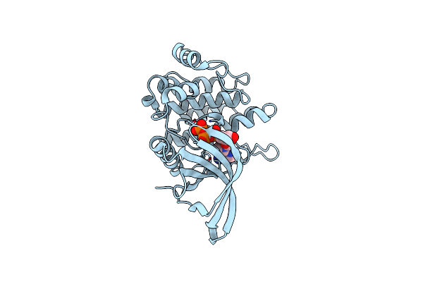
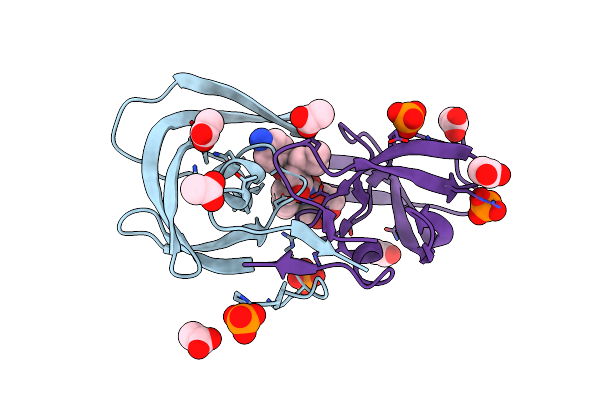
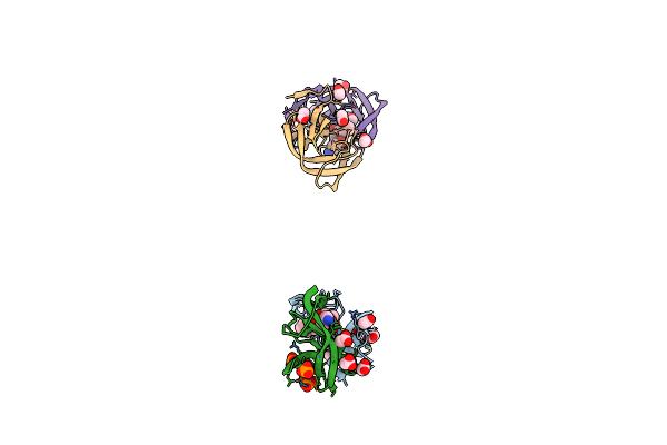
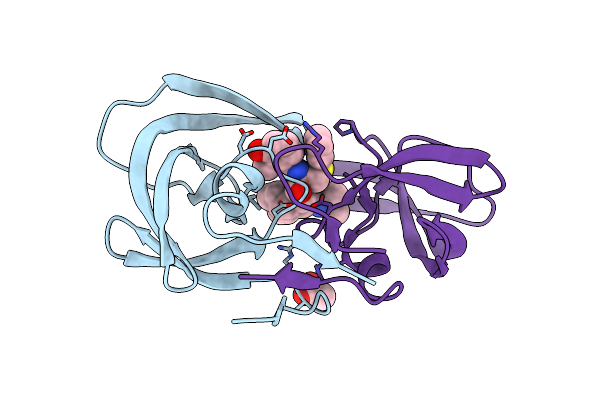
Mutations Outside The Active Site Of Hiv-1 Protease Alter Enzyme Structure And Dynamic Ensemble Of The Active Site To Confer Drug Resistance
Organism: Human immunodeficiency virus 1
Method: X-RAY DIFFRACTION Resolution:1.50 Å Release Date: 2015-02-18 Classification: hydrolase/hydrolase inhibitor Ligands: PO4, ACT, 017 |
 |
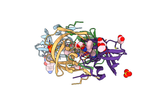
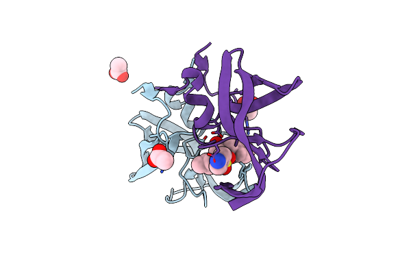
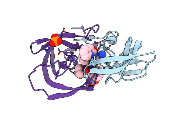
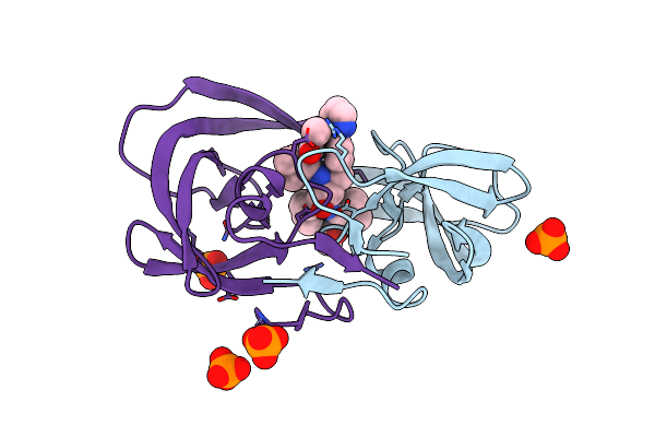
Organism: Homo sapiens
Method: X-RAY DIFFRACTION Resolution:2.01 Å Release Date: 2012-07-25 Classification: TRANSFERASE Ligands: GOL |
 |
Organism: Homo sapiens
Method: X-RAY DIFFRACTION Resolution:1.75 Å Release Date: 2012-07-25 Classification: TRANSFERASE Ligands: ATP, MG, ACT |
 |
Crystal Structure Of The Jak2 Pseudokinase Domain Mutant V617F (Mg-Atp-Bound Form)
Organism: Homo sapiens
Method: X-RAY DIFFRACTION Resolution:2.00 Å Release Date: 2012-07-25 Classification: TRANSFERASE Ligands: ATP, MG |
 |
Crystal Structure Of Hiv-1 I50V, A71 Protease In Complex With The Protease Inhibitor Amprenavir.
Organism: Hiv-1 m:b_arv2/sf2
Method: X-RAY DIFFRACTION Resolution:1.75 Å Release Date: 2011-09-28 Classification: Hydrolase/Hydrolase Inhibitor Ligands: 478, PO4, GOL, ACT, SO4 |
 |
Crystal Structure Of Hiv-1 I50V, A71V Protease In Complex With The Protease Inhibitor Darunavir
Organism: Hiv-1 m:b_arv2/sf2
Method: X-RAY DIFFRACTION Resolution:1.95 Å Release Date: 2011-09-28 Classification: Hydrolase/Hydrolase Inhibitor Ligands: 017, PO4, ACT, EDO, GOL |
 |
Crystal Structure Of Hiv-1 I50V, A71V Protease In Complex With The Protease Inhibitor Atazanavir
Organism: Hiv-1 m:b_arv2/sf2
Method: X-RAY DIFFRACTION Resolution:1.65 Å Release Date: 2011-09-28 Classification: Hydrolase/Hydrolase Inhibitor Ligands: GOL, EDO, ACT, DR7, PO4 |
 |
Crystal Structure Of Hiv-1 L76V Protease In Complex With The Protease Inhibitor Darunavir.
Organism: Human immunodeficiency virus type 1
Method: X-RAY DIFFRACTION Resolution:1.76 Å Release Date: 2011-02-09 Classification: Hydrolase/Hydrolase Inhibitor Ligands: PO4, ACT, 017 |
 |
Organism: Human immunodeficiency virus 1
Method: X-RAY DIFFRACTION Resolution:1.95 Å Release Date: 2010-08-11 Classification: HYDROLASE Ligands: 017, ACT |
 |
Crystal Structure Of A Nelfinavir Resistant Hiv-1 Crf01_Ae Protease Variant (N88S) In Complex With The Protease Inhibitor Darunavir.
Organism: Human immunodeficiency virus 1
Method: X-RAY DIFFRACTION Resolution:1.76 Å Release Date: 2010-08-11 Classification: HYDROLASE Ligands: ACT, 017 |
 |
Structure Of Nelfinavir-Resistant Hiv-1 Protease (D30N/N88D) In Complex With Darunavir.
Organism: Hiv-1 m:b_arv2/sf2
Method: X-RAY DIFFRACTION Resolution:2.15 Å Release Date: 2010-08-11 Classification: HYDROLASE Ligands: 017, PO4, ACT |
 |
Crystal Structure Of The Inhibitor Amprenavir (Apv) In Complex With A Multi-Drug Resistant Hiv-1 Protease Variant (L10I/G48V/I54V/V64I/V82A)Refer: Flap+ In Citation
Organism: Hiv-1 m:b_arv2/sf2
Method: X-RAY DIFFRACTION Resolution:2.15 Å Release Date: 2009-09-01 Classification: HYDROLASE Ligands: PO4, ACT, 478 |
 |
Crystal Structure Of Inhibitor Saquinavir (Sqv) In Complex With Multi-Drug Resistant Hiv-1 Protease (L63P/V82T/I84V) (Referred To As Act In Paper)
Organism: Hiv-1 m:b_arv2/sf2
Method: X-RAY DIFFRACTION Resolution:2.20 Å Release Date: 2009-09-01 Classification: HYDROLASE/HYDROLASE INHIBITOR Ligands: ROC, PO4 |
 |
Crystal Structure Of The Inhibitor Darunavir (Drv) In Complex With A Multi-Drug Resistant Hiv-1 Protease Variant (L10F/G48V/I54V/V64I/V82A) (Refer: Flap+ In Citation.)
Organism: Hiv-1 m:b_arv2/sf2
Method: X-RAY DIFFRACTION Resolution:1.97 Å Release Date: 2009-09-01 Classification: HYDROLASE Ligands: ACT, 017, PO4 |
 |
Crystal Structure Of The Wild Type Hiv-1 Protease With The Inhibitor, Amprenavir
Organism: Hiv-1 m:b_arv2/sf2
Method: X-RAY DIFFRACTION Resolution:1.75 Å Release Date: 2009-09-01 Classification: HYDROLASE Ligands: 478, ACT |
 |
Crystal Structure Of The Inhibitor Atazanavir (Atv) In Complex With A Multi-Drug Resistance Hiv-1 Protease Variant (L10I/G48V/I54V/V64I/V82A) Refer: Flap+ In Citation.
Organism: Hiv-1 m:b_arv2/sf2
Method: X-RAY DIFFRACTION Resolution:1.60 Å Release Date: 2009-09-01 Classification: HYDROLASE Ligands: PO4, DR7 |
 |
Crystal Structure Of The Wild-Type Hiv-1 Protease With The Inhibitor, Nelfinavir
Organism: Hiv-1 m:b_arv2/sf2
Method: X-RAY DIFFRACTION Resolution:1.97 Å Release Date: 2009-09-01 Classification: HYDROLASE Ligands: 1UN, ACT |
 |
Crystal Structure Of Wild-Type Hiv Protease In Complex With The Inhibitor, Atazanavir
Organism: Hiv-1 m:b_arv2/sf2
Method: X-RAY DIFFRACTION Resolution:1.80 Å Release Date: 2009-09-01 Classification: HYDROLASE Ligands: DR7, PO4 |