
Search Count: 14
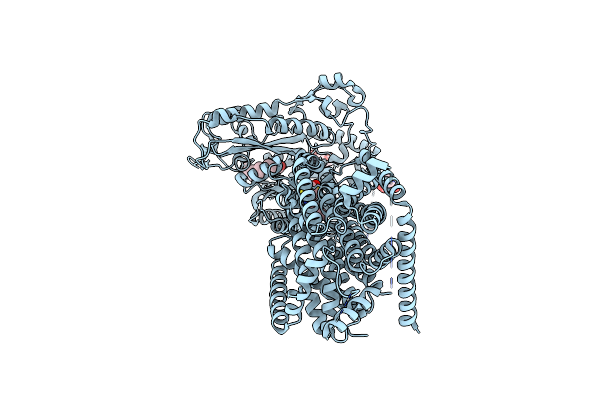 |
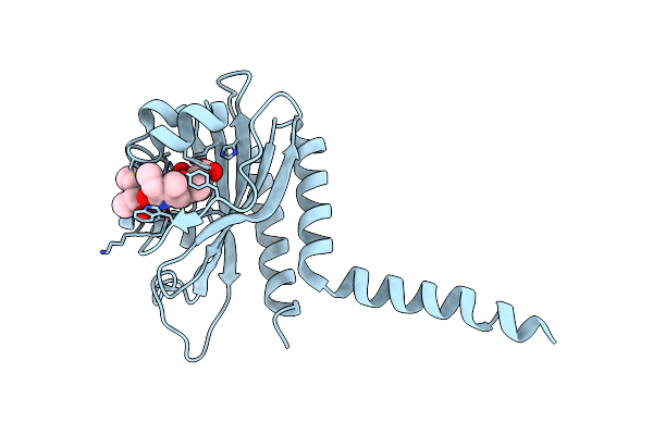
Plasmodium Falciparum Niemann-Pick Type C1-Related Protein Bound With Mmv009108
Organism: Plasmodium falciparum 3d7
Method: ELECTRON MICROSCOPY Release Date: 2024-11-20 Classification: MEMBRANE PROTEIN Ligands: NAG, CLR, Y6F |
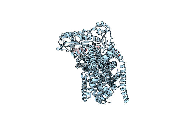 |
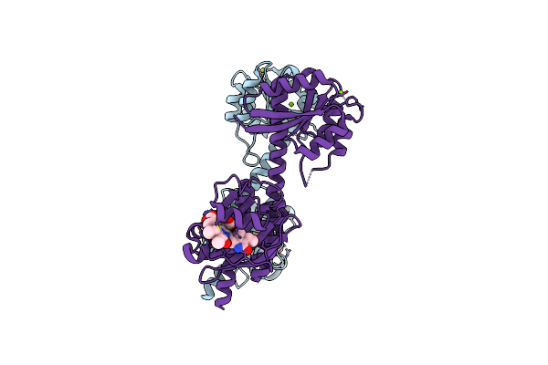
Organism: Plasmodium falciparum 3d7
Method: ELECTRON MICROSCOPY Release Date: 2024-11-13 Classification: MEMBRANE PROTEIN Ligands: CLR, NAG |
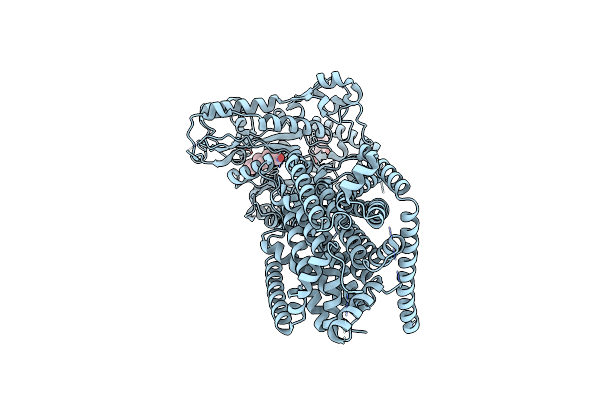 |
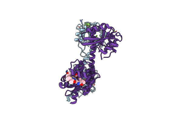
Organism: Plasmodium falciparum 3d7
Method: ELECTRON MICROSCOPY Release Date: 2024-11-13 Classification: MEMBRANE PROTEIN Ligands: CLR, NAG |
 |
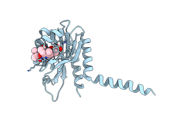
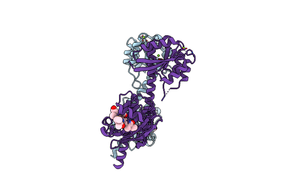
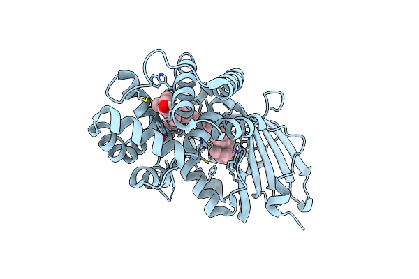
Crystal Structure Of The Histidine Kinase Domain Of Bacteriophytochrome Rpbphp2
Organism: Rhodopseudomonas palustris cga009
Method: X-RAY DIFFRACTION Resolution:3.19 Å Release Date: 2023-11-22 Classification: SIGNALING PROTEIN, TRANSFERASE Ligands: ATP, MG |
 |
Crystal Structure Of A Far-Red Cyanobacteriochrome Photoreceptor At Room Temperature
Organism: Anabaena cylindrica (strain atcc 27899 / pcc 7122)
Method: X-RAY DIFFRACTION Resolution:2.70 Å Release Date: 2020-11-04 Classification: SIGNALING PROTEIN Ligands: CYC |
 |
Organism: Anabaena cylindrica (strain atcc 27899 / pcc 7122)
Method: X-RAY DIFFRACTION Resolution:3.00 Å Release Date: 2020-11-04 Classification: SIGNALING PROTEIN Ligands: CYC |
 |
Crystal Structure Of A Dual Sensor Histidine Kinase In The Green-Light Absorbing Pg State
Organism: [leptolyngbya] sp. jsc-1
Method: X-RAY DIFFRACTION Resolution:1.97 Å Release Date: 2019-09-18 Classification: SIGNALING PROTEIN Ligands: CYC, MG |
 |
Organism: [leptolyngbya] sp. jsc-1
Method: X-RAY DIFFRACTION Resolution:2.54 Å Release Date: 2019-09-18 Classification: SIGNALING PROTEIN Ligands: BEF, CYC, MG |
 |
Crystal Structure Of A Dual Sensor Histidine Kinase In Green Light Illuminated State
Organism: [leptolyngbya] sp. jsc-1
Method: X-RAY DIFFRACTION Resolution:2.30 Å Release Date: 2019-09-18 Classification: SIGNALING PROTEIN Ligands: CYC, MG |
 |
Crystal Structure Of Bacterial (6-4) Photolyase Phrb From In Situ Serial Laue Diffraction
Organism: Rhizobium radiobacter
Method: X-RAY DIFFRACTION Resolution:2.30 Å Release Date: 2018-07-18 Classification: LYASE Ligands: FAD, DLZ, SF4 |
 |
Crystal Structure Of Plant Uvb Photoreceptor Uvr8 From In Situ Serial Laue Diffraction
Organism: Arabidopsis thaliana
Method: X-RAY DIFFRACTION Resolution:2.00 Å Release Date: 2018-07-18 Classification: GENE REGULATION |
 |
Crystal Structure Of Orange Carotenoid Protein With Partial Loss Of 3'Oh Echinenone Chromophore
Organism: Synechocystis sp. (strain pcc 6803 / kazusa)
Method: X-RAY DIFFRACTION Resolution:2.30 Å Release Date: 2017-06-07 Classification: Carotenoid binding protein Ligands: GOL, EQ3 |
 |
Crystal Structure And Light Induced Structural Changes In Orange Carotenoid Protein Bound With Echinenone
Organism: Synechocystis sp. (strain pcc 6803 / kazusa)
Method: X-RAY DIFFRACTION Resolution:1.50 Å Release Date: 2017-06-07 Classification: Carotenoid binding protein Ligands: ECH, GOL |
 |
Crystal Structure And Light Induced Structural Changes In Orange Carotenoid Protein Bound With 3 'Oh Echinenone
Organism: Synechocystis sp. (strain pcc 6803 / kazusa)
Method: X-RAY DIFFRACTION Resolution:1.65 Å Release Date: 2017-06-07 Classification: Carotenoid binding protein Ligands: GOL, EQ3 |