
Search Count: 24
 |
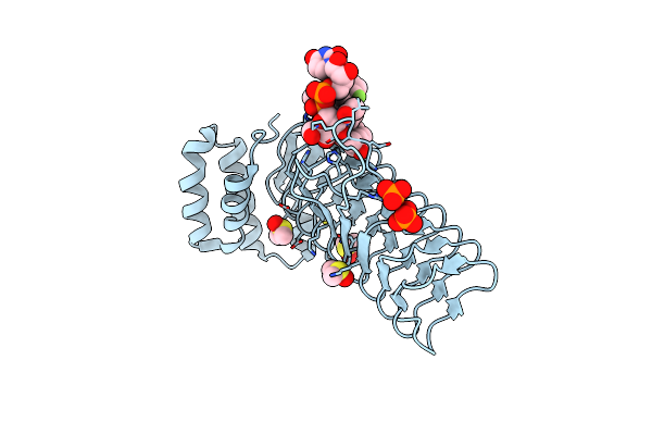
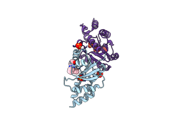
Organism: Homo sapiens
Method: X-RAY DIFFRACTION Resolution:1.91 Å Release Date: 2024-09-04 Classification: ANTIBIOTIC Ligands: MYR, A1AZR |
 |
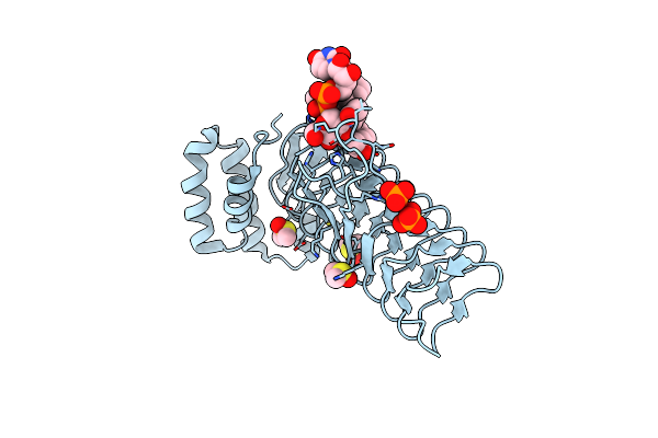
Organism: Acinetobacter baumannii
Method: X-RAY DIFFRACTION Resolution:2.80 Å Release Date: 2024-09-04 Classification: ANTIBIOTIC Ligands: ANP, MVC, MG, A1AZS |
 |
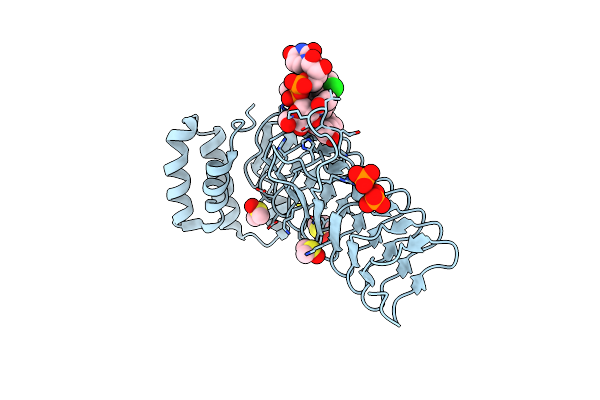
Organism: Acinetobacter baumannii
Method: ELECTRON MICROSCOPY Release Date: 2024-04-24 Classification: ANTIMICROBIAL PROTEIN Ligands: ANP, ZQF |
 |
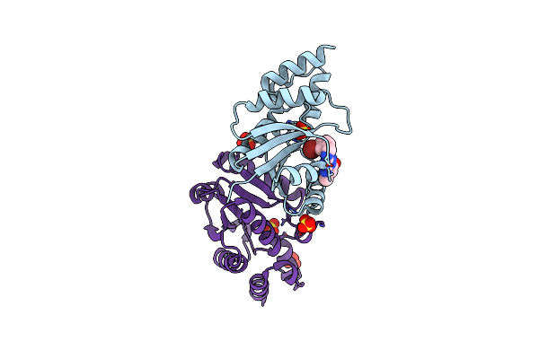
Organism: Human immunodeficiency virus type 1
Method: X-RAY DIFFRACTION Resolution:2.18 Å Release Date: 2022-11-23 Classification: VIRAL PROTEIN Ligands: 9QI |
 |
Organism: Human immunodeficiency virus type 1
Method: X-RAY DIFFRACTION Resolution:2.08 Å Release Date: 2022-11-23 Classification: VIRAL PROTEIN Ligands: 9PJ |
 |
Organism: Homo sapiens
Method: X-RAY DIFFRACTION Resolution:2.46 Å Release Date: 2022-10-05 Classification: HYDROLASE Ligands: D06 |
 |
Organism: Homo sapiens
Method: X-RAY DIFFRACTION Resolution:2.69 Å Release Date: 2022-10-05 Classification: HYDROLASE Ligands: D06, TMO |
 |
Organism: Homo sapiens
Method: X-RAY DIFFRACTION Resolution:2.78 Å Release Date: 2022-10-05 Classification: HYDROLASE Ligands: CW8 |
 |
Organism: Homo sapiens
Method: X-RAY DIFFRACTION Resolution:2.58 Å Release Date: 2022-10-05 Classification: HYDROLASE Ligands: CW8, TMO |
 |
K. Pneumoniae Topoisomerase Iv (Pare-Parc) In Complex With Dna And Compound 34 (7-[(1S,5R)-1-Amino-3-Azabicyclo[3.1.0]Hexan-3-Yl]-4-(Aminomethyl)-1-Cyclopropyl-3,6-Difluoro-8-Methylquinolin-2(1H)-One)
Organism: Klebsiella pneumoniae 342
Method: X-RAY DIFFRACTION Resolution:3.20 Å Release Date: 2020-07-15 Classification: ISOMERASE/DNA/ISOMERASE INHIBITOR Ligands: MPD, MG, CL, TNJ |
 |
Organism: Escherichia coli
Method: X-RAY DIFFRACTION Resolution:2.00 Å Release Date: 2020-03-11 Classification: TRANSFERASE/INHIBITOR Ligands: PO4, DMS, O5J |
 |
E.Coli Lpxa In Complex With Udp-3-O-(R-3-Hydroxymyristoyl)-Glcnac And Compound 2
Organism: Escherichia coli
Method: X-RAY DIFFRACTION Resolution:1.70 Å Release Date: 2020-03-11 Classification: TRANSFERASE/INHIBITOR Ligands: U20, PO4, DMS, O5P |
 |
E.Coli Lpxa In Complex With Udp-3-O-(R-3-Hydroxymyristoyl)-Glcnac And Compound 6
Organism: Escherichia coli
Method: X-RAY DIFFRACTION Resolution:1.75 Å Release Date: 2020-03-11 Classification: TRANSFERASE/INHIBITOR Ligands: U20, PO4, DMS, O5M |
 |
E.Coli Lpxa In Complex With Udp-3-O-(R-3-Hydroxymyristoyl)-Glcnac And Compound 7
Organism: Escherichia coli
Method: X-RAY DIFFRACTION Resolution:1.70 Å Release Date: 2020-03-11 Classification: TRANSFERASE/INHIBITOR Ligands: U20, PO4, DMS, O5G |
 |
E.Coli Lpxa In Complex With Udp-3-O-(R-3-Hydroxymyristoyl)-Glcnac And Compound 8
Organism: Escherichia coli
Method: X-RAY DIFFRACTION Resolution:1.75 Å Release Date: 2020-03-11 Classification: TRANSFERASE/INHIBITOR Ligands: U20, PO4, DMS, O5D |
 |
Crystal Structure Of E.Coli Phosphopantetheine Adenylyltransferase (Ppat/Coad) In Complex With (R)-3-(3-Chlorophenyl)-3-((5-Methyl-7-Oxo-4,7-Dihydro-[1,2,4]Triazolo[1,5-A]Pyrimidin-2-Yl)Amino)Propanenitrile
Organism: Escherichia coli (strain k12)
Method: X-RAY DIFFRACTION Resolution:1.61 Å Release Date: 2018-03-14 Classification: TRANSFERASE/antibiotic Ligands: EXJ, ATP, MG, SO4 |
 |
Crystal Structure Of E.Coli Phosphopantetheine Adenylyltransferase (Ppat/Coad) In Complex With 1-Benzyl-1H-Imidazo[4,5-B]Pyridine
Organism: Escherichia coli (strain k12)
Method: X-RAY DIFFRACTION Resolution:1.77 Å Release Date: 2018-03-14 Classification: TRANSFERASE/antibiotic Ligands: EXG, SO4, DMS |
 |
Crystal Structure Of E.Coli Phosphopantetheine Adenylyltransferase (Ppat/Coad) In Complex With 2-((3-Bromobenzyl)Amino)-5-Methyl-[1,2,4]Triazolo[1,5-A]Pyrimidin-7(4H)-One
Organism: Escherichia coli (strain k12)
Method: X-RAY DIFFRACTION Resolution:1.79 Å Release Date: 2018-03-14 Classification: TRANSFERASE/antibiotic Ligands: EXP, SO4 |
 |
Crystal Structure Of E.Coli Phosphopantetheine Adenylyltransferase (Ppat/Coad) In Complex With (R)-2,4-Dihydroxy-N-(2-(4-Hydroxy-1H-Benzo[D]Imidazol-2-Yl)Ethyl)-3,3-Dimethylbutanamide
Organism: Escherichia coli (strain k12)
Method: X-RAY DIFFRACTION Resolution:1.87 Å Release Date: 2018-03-14 Classification: TRANSFERASE/antibiotic Ligands: EXS, SO4 |
 |
Crystal Structure Of E.Coli Phosphopantetheine Adenylyltransferase (Ppat/Coad) In Complex With 3-((1S,2S)-2-(4-Hydroxy-1H-Benzo[D]Imidazol-2-Yl)Cyclopentyl)Benzoic Acid
Organism: Escherichia coli (strain k12)
Method: X-RAY DIFFRACTION Resolution:1.82 Å Release Date: 2018-03-14 Classification: TRANSFERASE/antibiotic Ligands: EXV, SO4 |