
Search Count: 74
 |
Organism: Escherichia coli
Method: ELECTRON MICROSCOPY Release Date: 2025-11-26 Classification: HYDROLASE Ligands: MG, NA |
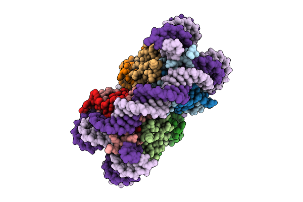 |
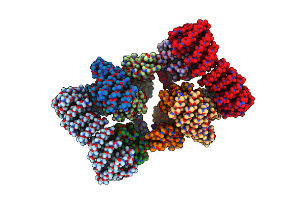
Human Nucleosome Structure On Nickel-Nta Lipid Affinity Grid (C2 Refinement)
Organism: Homo sapiens
Method: ELECTRON MICROSCOPY Resolution:2.59 Å Release Date: 2025-10-29 Classification: DNA BINDING PROTEIN |
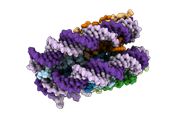 |
Human Nucleosome Structure On Nickel-Nta Lipid Affinity Grid (C1 Refinement)
Organism: Homo sapiens
Method: ELECTRON MICROSCOPY Resolution:2.74 Å Release Date: 2025-10-29 Classification: DNA BINDING PROTEIN |
 |
Organism: Saccharomyces cerevisiae
Method: ELECTRON MICROSCOPY Resolution:3.10 Å Release Date: 2025-10-29 Classification: EXOCYTOSIS |
 |
Organism: Staphylococcus aureus
Method: X-RAY DIFFRACTION Resolution:1.75 Å Release Date: 2025-10-01 Classification: CELL CYCLE |
 |
Organism: Homo sapiens
Method: ELECTRON MICROSCOPY Release Date: 2024-12-18 Classification: TRANSPORT PROTEIN Ligands: MG, GTP |
 |
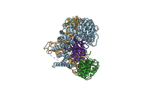
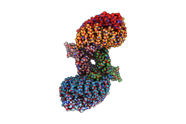
Human Ap-3 Dimer Bound To Myristoylated Arf1 (Q71L) And Lamp1 Cargo On A Lipid Nanodisc
Organism: Homo sapiens
Method: ELECTRON MICROSCOPY Release Date: 2024-12-18 Classification: TRANSPORT PROTEIN Ligands: MG, GTP |
 |
Organism: Homo sapiens
Method: ELECTRON MICROSCOPY Release Date: 2024-12-18 Classification: TRANSPORT PROTEIN Ligands: MG, GTP |
 |
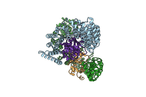
Ap-3 Bound To Myristoylated Arf1 (Q71L) And Lampi On A Lipid Nanodisc; Combined Map
Organism: Homo sapiens
Method: ELECTRON MICROSCOPY Release Date: 2024-12-18 Classification: TRANSPORT PROTEIN Ligands: MG, GTP |
 |
Organism: Homo sapiens
Method: ELECTRON MICROSCOPY Release Date: 2024-12-18 Classification: TRANSPORT PROTEIN |
 |
Organism: Mus musculus, Rattus norvegicus
Method: ELECTRON MICROSCOPY Release Date: 2023-07-12 Classification: ENDOCYTOSIS Ligands: PIO |
 |
Structure Of Hdac6 Zinc-Finger Ubiquitin Binding Domain In Complex With 3-(3-(2-(Methylamino)-2-Oxoethyl)-4-Oxo-3,4-Dihydroquinazolin-2-Yl)Propanoic Acid
Organism: Homo sapiens
Method: X-RAY DIFFRACTION Resolution:1.55 Å Release Date: 2023-05-03 Classification: HYDROLASE/INHIBITOR Ligands: ZN, ZU6 |
 |
Structure Of Hdac6 Zinc-Finger Ubiquitin Binding Domain In Complex With 3-(3-(2-(Benzylamino)-2-Oxoethyl)-4-Oxo-3,4-Dihydroquinazolin-2-Yl)Propanoic Acid
Organism: Homo sapiens
Method: X-RAY DIFFRACTION Resolution:1.55 Å Release Date: 2023-05-03 Classification: HYDROLASE/INHIBITOR Ligands: ZN, ZU9 |
 |
Structure Of Hdac6 Zinc-Finger Ubiquitin Binding Domain In Complex With Sgc-Ubd253 Chemical Probe
Organism: Homo sapiens
Method: X-RAY DIFFRACTION Resolution:1.62 Å Release Date: 2023-05-03 Classification: HYDROLASE/INHIBITOR Ligands: ZN, ZUE |
 |
Organism: Saccharomyces cerevisiae
Method: X-RAY DIFFRACTION Resolution:2.22 Å Release Date: 2022-05-25 Classification: TRANSPORT PROTEIN |
 |
Organism: Mus musculus
Method: ELECTRON MICROSCOPY Release Date: 2022-03-30 Classification: ENDOCYTOSIS |
 |
Organism: Mus musculus
Method: ELECTRON MICROSCOPY Release Date: 2022-03-30 Classification: ENDOCYTOSIS |
 |
Organism: Mus musculus
Method: ELECTRON MICROSCOPY Release Date: 2022-03-30 Classification: ENDOCYTOSIS |
 |
Organism: Mus musculus
Method: ELECTRON MICROSCOPY Release Date: 2022-03-30 Classification: ENDOCYTOSIS |
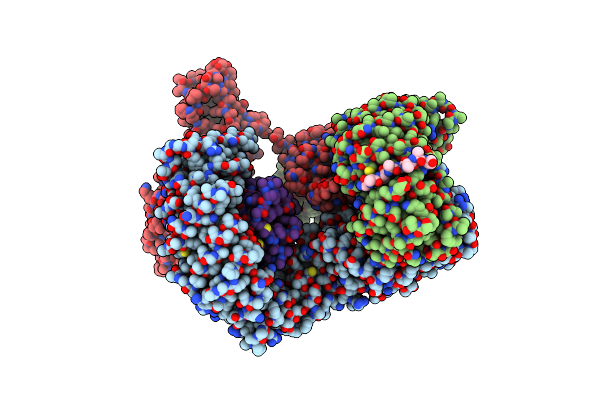 |
Ap2 Bound To The Apa Domain Of Sgip And Heparin; Partial Signal Subtraction And Symmetry Expansion
Organism: Mus musculus
Method: ELECTRON MICROSCOPY Release Date: 2022-03-30 Classification: ENDOCYTOSIS |