
Search Count: 25
 |
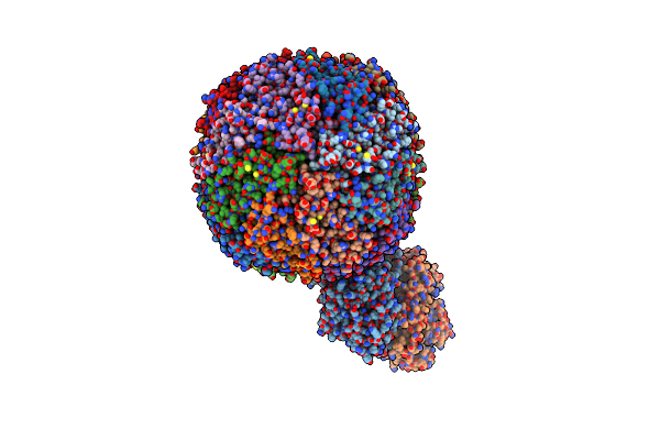
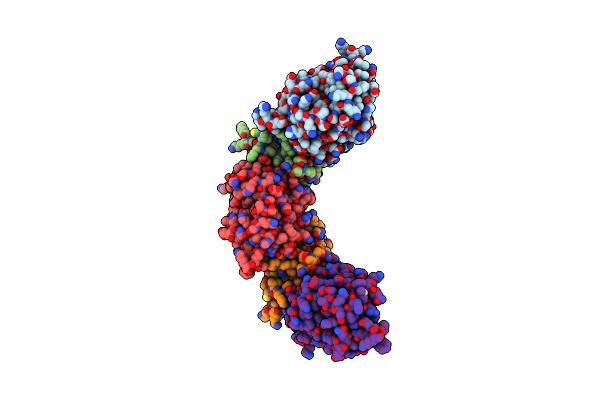
Single Particle Cryo-Em Map Of Human Transferrin Receptor 1 - H-Ferritin Complex At 5.5 Angstrom Resolution.
Organism: Homo sapiens
Method: ELECTRON MICROSCOPY Release Date: 2019-03-27 Classification: METAL BINDING PROTEIN |
 |
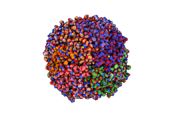
Single Particle Cryo-Em Map Of Human Transferrin Receptor 1 - H-Ferritin Complex.
Organism: Homo sapiens
Method: ELECTRON MICROSCOPY Release Date: 2019-03-27 Classification: METAL BINDING PROTEIN |
 |
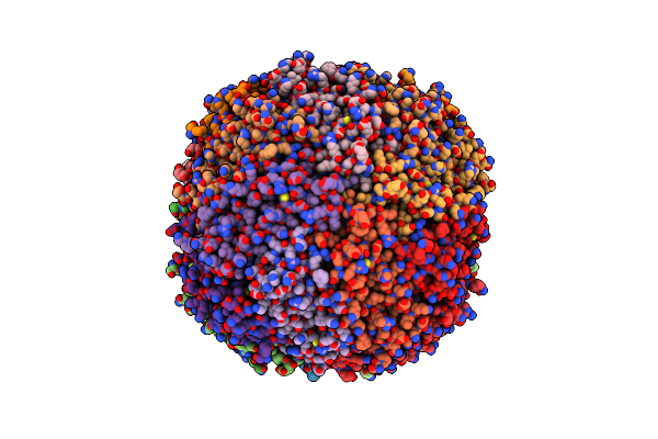
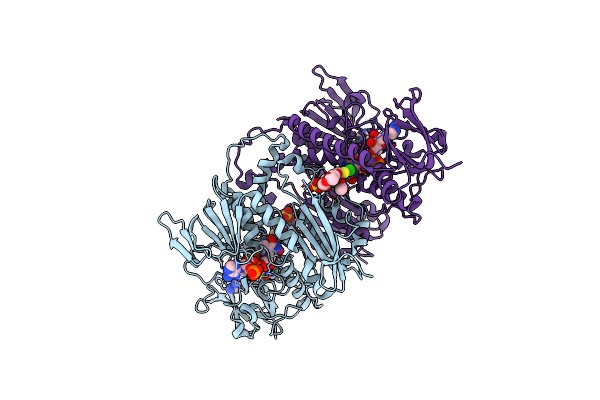
Structure Of A Modified Mouse H-Chain Ferritin With A Lanthanide Binding Motif
Organism: Mus musculus
Method: X-RAY DIFFRACTION Resolution:2.85 Å Release Date: 2018-07-25 Classification: METAL TRANSPORT Ligands: FE |
 |
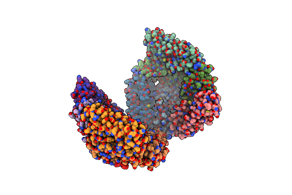
Structure Of A Modified Mouse H Chain Ferritin With A Lanthanide Binding Motif In Complex With Terbium
Organism: Mus musculus
Method: X-RAY DIFFRACTION Resolution:2.65 Å Release Date: 2018-07-25 Classification: METAL TRANSPORT Ligands: TB |
 |
Organism: Archaeoglobus fulgidus
Method: X-RAY DIFFRACTION Resolution:2.93 Å Release Date: 2016-11-30 Classification: METAL BINDING PROTEIN Ligands: MG |
 |
Organism: Thermobifida fusca tm51
Method: X-RAY DIFFRACTION Resolution:3.40 Å Release Date: 2014-09-17 Classification: OXIDOREDUCTASE Ligands: HEM, ACT |
 |
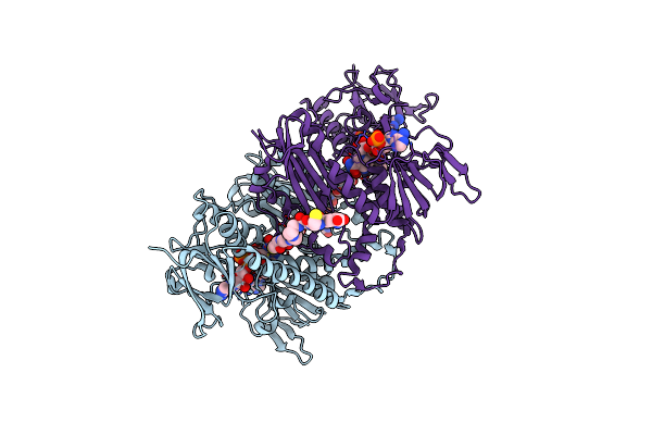
Structure Of Tr From Leishmania Infantum In Complex With A Diarylpirrole-Based Inhibitor
Organism: Leishmania infantum
Method: X-RAY DIFFRACTION Resolution:3.20 Å Release Date: 2013-04-17 Classification: OXIDOREDUCTASE Ligands: FAD, NDP, JV0 |
 |
Crystal Structure Of Leishmania Infantum Trypanothione Reductase In Complex With Nadph And Trypanothione
Organism: Leishmania infantum
Method: X-RAY DIFFRACTION Resolution:3.61 Å Release Date: 2013-01-16 Classification: OXIDOREDUCTASE Ligands: FAD, NDP, GCG |
 |
Organism: Leishmania major
Method: X-RAY DIFFRACTION Resolution:3.00 Å Release Date: 2012-09-05 Classification: OXIDOREDUCTASE |
 |
Organism: Leishmania major
Method: X-RAY DIFFRACTION Resolution:1.80 Å Release Date: 2012-06-13 Classification: ELECTRON TRANSPORT Ligands: MG |
 |
X-Ray Structure Of The Leishmania Infantum Tryopanothione Reductase In Complex With Auranofin
Organism: Leishmania infantum
Method: X-RAY DIFFRACTION Resolution:3.50 Å Release Date: 2012-01-11 Classification: OXIDOREDUCTASE Ligands: FAD, NDP, AU, TS8, CL, SO4 |
 |
Crystal Structure Of Trypanothione Reductase From Leishmania Infantum In Complex With Nadph And Silver
Organism: Leishmania infantum
Method: X-RAY DIFFRACTION Resolution:3.30 Å Release Date: 2011-01-12 Classification: OXIDOREDUCTASE Ligands: FAD, NDP, AG, SO4 |
 |
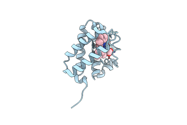
Structure Of Mk-3281, A Potent Non-Nucleoside Finger-Loop Inhibitor, In Complex With The Hepatitis C Virus Ns5B Polymerase
Organism: Hepatitis c virus
Method: X-RAY DIFFRACTION Resolution:2.53 Å Release Date: 2010-12-22 Classification: TRANSFERASE Ligands: IB8, MN |
 |
Organism: Leishmania infantum
Method: X-RAY DIFFRACTION Resolution:2.95 Å Release Date: 2009-04-28 Classification: OXIDOREDUCTASE Ligands: FAD, SO4 |
 |
X Ray Structure Of Leishmania Infantum Trypanothione Reductase In Complex With Antimony And Nadph
Organism: Leishmania infantum
Method: X-RAY DIFFRACTION Resolution:3.00 Å Release Date: 2009-04-28 Classification: OXIDOREDUCTASE Ligands: FAD, SB, NDP, SO4 |
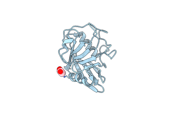 |
Organism: Schistosoma mansoni
Method: X-RAY DIFFRACTION Resolution:1.80 Å Release Date: 2007-05-15 Classification: ISOMERASE Ligands: ACT |
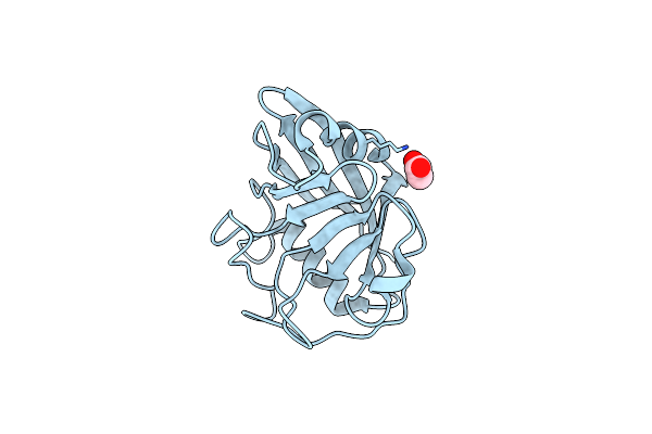 |
Organism: Schistosoma mansoni
Method: X-RAY DIFFRACTION Resolution:1.50 Å Release Date: 2007-05-15 Classification: ISOMERASE Ligands: ACT |
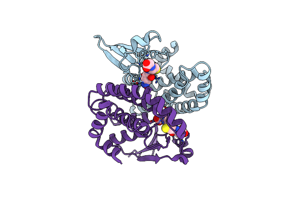 |
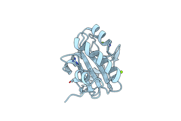
Crystal Structure Of The Y10F Mutant Of The Gluathione S-Transferase From Schistosoma Haematobium
Organism: Schistosoma haematobium
Method: X-RAY DIFFRACTION Resolution:2.10 Å Release Date: 2006-07-04 Classification: TRANSFERASE Ligands: GSH |
 |
Organism: Schistosoma haematobium
Method: X-RAY DIFFRACTION Resolution:2.49 Å Release Date: 2006-06-26 Classification: TRANSFERASE Ligands: GSH, PG4 |
 |
Organism: Schistosoma haematobium
Method: X-RAY DIFFRACTION Resolution:2.30 Å Release Date: 2006-06-21 Classification: TRANSFERASE Ligands: GTX, PG4 |