
Search Count: 80
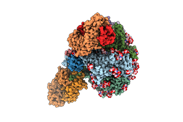 |
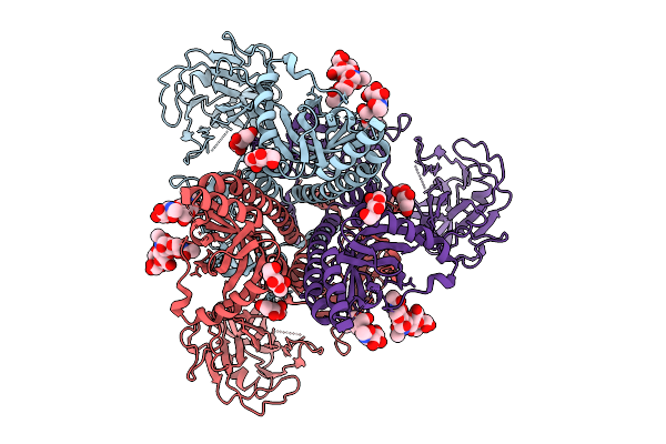
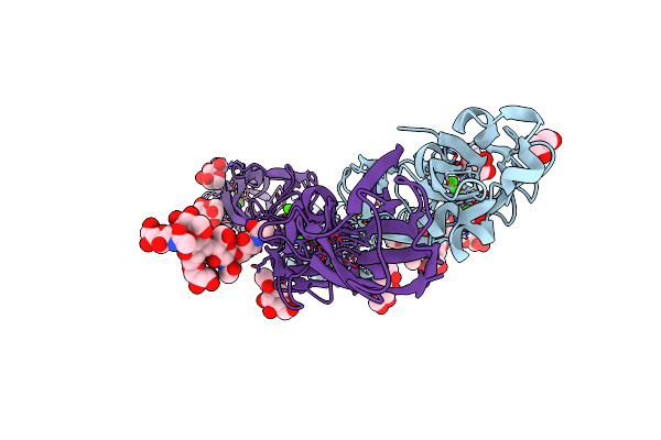
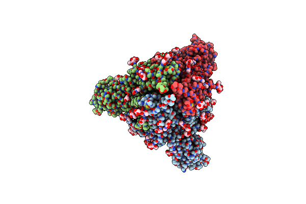
Cryo-Em Structure Of Hiv-1 Bg505Ds-Sosip.664 Env Trimer Bound To Dfph-A.01_10R59P_Lc Fab
Organism: Human immunodeficiency virus 1, Macaca
Method: ELECTRON MICROSCOPY Release Date: 2025-12-10 Classification: VIRAL PROTEIN Ligands: NAG |
 |
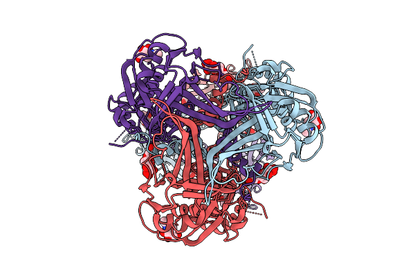
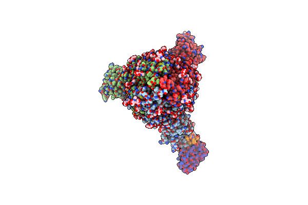
Cryo-Em Structure Of Neutralizing Murine Antibody Ws.Hsv-1.24 In Complex With Hsv-1 Glycoprotein B Trimer Gb-Ecto.516P.531E
Organism: Human alphaherpesvirus 1, Mus musculus
Method: ELECTRON MICROSCOPY Release Date: 2025-09-10 Classification: VIRAL PROTEIN Ligands: NAG |
 |
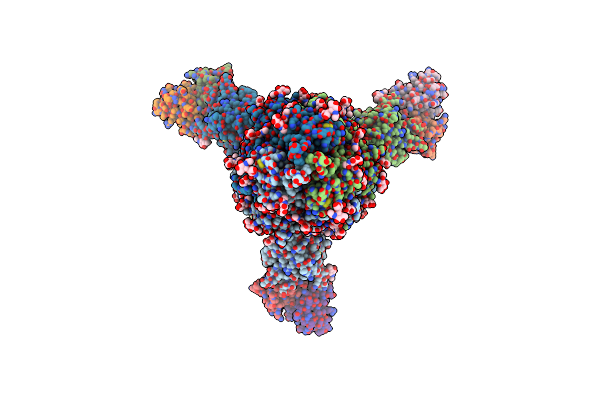
Cryo-Em Structure Of Neutralizing Human Antibody D48 In Complex With Hsv-1 Glycoprotein B Trimer Gb-Ecto.516P.531E.Ds
Organism: Human alphaherpesvirus 1, Homo sapiens
Method: ELECTRON MICROSCOPY Release Date: 2025-09-10 Classification: VIRAL PROTEIN Ligands: NAG |
 |
Cryo-Em Structure Of Neutralizing Human Antibody D48 In Complex With Hsv-1 Glycoprotein B Trimer Gb-Ecto.516P
Organism: Human alphaherpesvirus 1, Homo sapiens
Method: ELECTRON MICROSCOPY Release Date: 2025-09-10 Classification: VIRAL PROTEIN Ligands: NAG |
 |
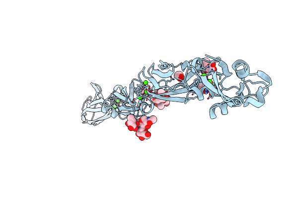
Cryo-Em Structure Of Gb-Ecto.516P.531E.Ds, A Prefusion-Stabilized Hsv-1 Glycoprotein B Extracellular Domain
Organism: Human alphaherpesvirus 1
Method: ELECTRON MICROSCOPY Release Date: 2025-09-10 Classification: VIRAL PROTEIN Ligands: NAG |
 |
Cryo-Em Structure Of Gb-Ecto.516P, An Hsv-1 Glycoprotein B Extracellular Domain
Organism: Human alphaherpesvirus 1
Method: ELECTRON MICROSCOPY Release Date: 2025-09-10 Classification: VIRAL PROTEIN Ligands: NAG |
 |
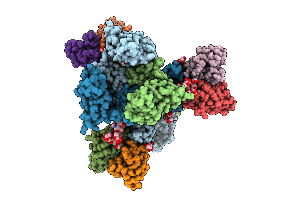
Organism: Drosophila melanogaster
Method: ELECTRON MICROSCOPY Release Date: 2025-01-29 Classification: SIGNALING PROTEIN Ligands: NAG |
 |
Organism: Drosophila melanogaster
Method: ELECTRON MICROSCOPY Release Date: 2025-01-29 Classification: SIGNALING PROTEIN Ligands: NAG |
 |
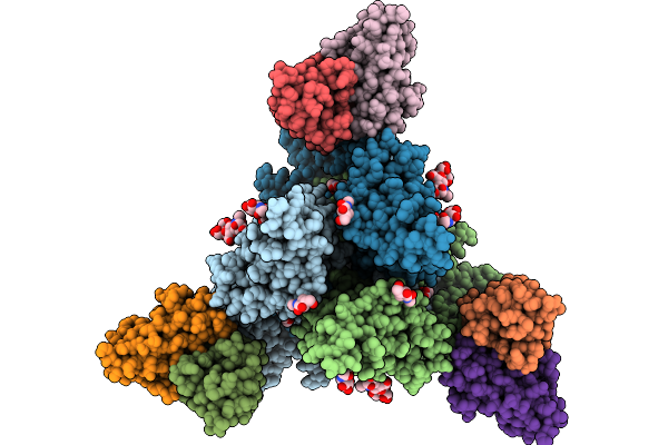
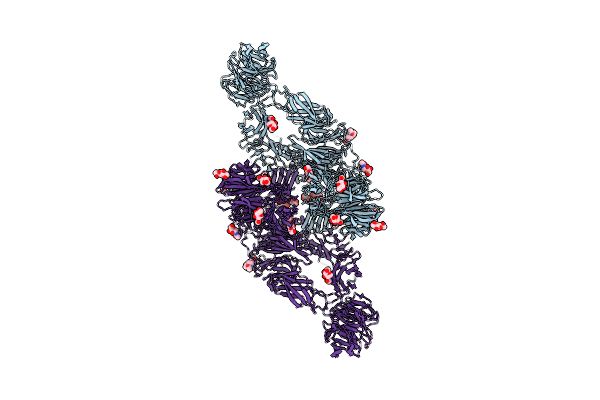
Cryo-Em Structure Of Bride Of Sevenless Extracellular Domain (Dimer, Sevenless-Bound Form)
Organism: Drosophila melanogaster
Method: ELECTRON MICROSCOPY Release Date: 2025-01-29 Classification: SIGNALING PROTEIN Ligands: NAG |
 |
Organism: Drosophila melanogaster
Method: ELECTRON MICROSCOPY Release Date: 2025-01-29 Classification: SIGNALING PROTEIN Ligands: NAG |
 |
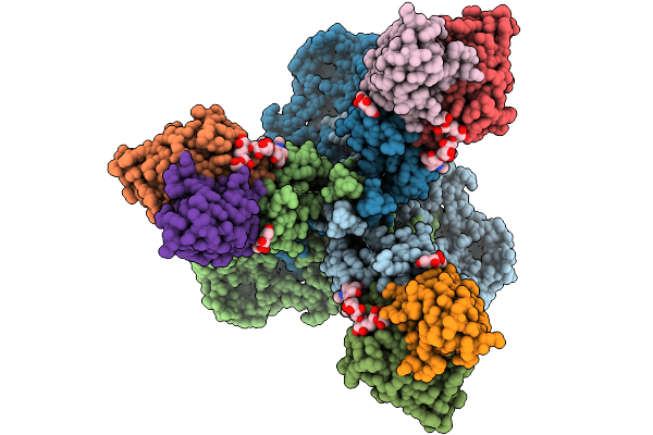
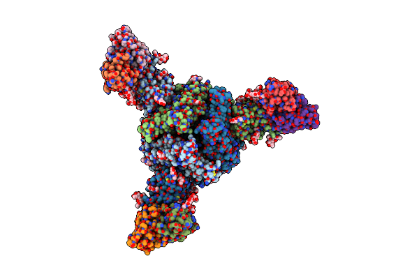
Cryo-Em Structure Of Sevenless Extracellular Domain (Composite Map Of The Dimer, Ph 4.6)
Organism: Drosophila melanogaster
Method: ELECTRON MICROSCOPY Release Date: 2025-01-29 Classification: SIGNALING PROTEIN Ligands: NAG |
 |
Cryo-Em Structure Of Ntd-Directed Neutralizing Antibody Lp5 Fab In Complex With Sars-Cov-2 S2P Spike
Organism: Severe acute respiratory syndrome coronavirus 2, Homo sapiens
Method: ELECTRON MICROSCOPY Release Date: 2022-05-25 Classification: VIRAL PROTEIN/IMMUNE SYSTEM Ligands: NAG |
 |
Organism: Mus musculus
Method: X-RAY DIFFRACTION Resolution:2.40 Å Release Date: 2022-03-16 Classification: CELL ADHESION Ligands: MAN, NAG, GOL, CA, CL |
 |
Cryo-Em Structure Of Ntd-Directed Neutralizing Antibody 5-7 In Complex With Prefusion Sars-Cov-2 Spike Glycoprotein
Organism: Severe acute respiratory syndrome coronavirus 2, Homo sapiens
Method: ELECTRON MICROSCOPY Release Date: 2021-09-01 Classification: VIRAL PROTEIN/IMMUNE SYSTEM Ligands: NAG |
 |
Organism: Mus musculus
Method: X-RAY DIFFRACTION Resolution:3.51 Å Release Date: 2021-07-14 Classification: CELL ADHESION Ligands: MAN, CA, EDO |
 |
Organism: Severe acute respiratory syndrome coronavirus 2, Homo sapiens
Method: ELECTRON MICROSCOPY Release Date: 2021-04-14 Classification: VIRAL PROTEIN/Immune System Ligands: NAG |
 |
Organism: Severe acute respiratory syndrome coronavirus 2, Homo sapiens
Method: ELECTRON MICROSCOPY Release Date: 2021-04-14 Classification: VIRAL PROTEIN/Immune System Ligands: NAG |
 |
Organism: Severe acute respiratory syndrome coronavirus 2, Homo sapiens
Method: ELECTRON MICROSCOPY Release Date: 2021-04-14 Classification: VIRAL PROTEIN/Immune System Ligands: NAG |
 |
Cryo-Em Structure Of Ntd-Directed Neutralizing Antibody 1-87 In Complex With Prefusion Sars-Cov-2 Spike Glycoprotein
Organism: Severe acute respiratory syndrome coronavirus 2, Homo sapiens
Method: ELECTRON MICROSCOPY Release Date: 2021-03-24 Classification: Viral protein/Immune system Ligands: NAG |
 |
Cryo-Em Structure Of Ntd-Directed Neutralizing Antibody 4-18 In Complex With Prefusion Sars-Cov-2 Spike Glycoprotein
Organism: Severe acute respiratory syndrome coronavirus 2, Homo sapiens
Method: ELECTRON MICROSCOPY Release Date: 2021-03-24 Classification: Viral Protein/IMMUNE SYSTEM Ligands: NAG |