
Search Count: 14
 |
Organism: Homo sapiens, Mus musculus
Method: ELECTRON MICROSCOPY Release Date: 2025-03-19 Classification: SIGNALING PROTEIN |
 |
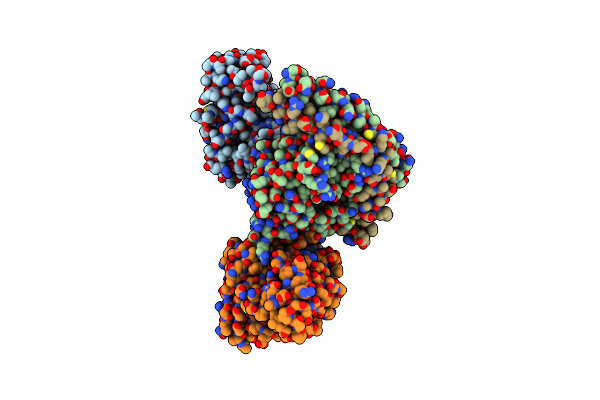
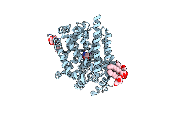
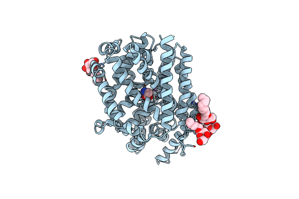
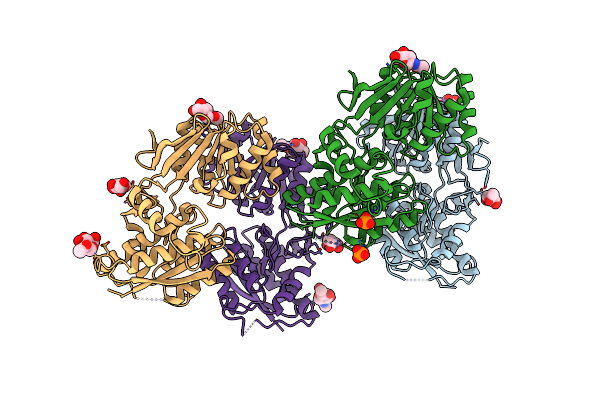
Organism: Homo sapiens
Method: X-RAY DIFFRACTION Resolution:2.70 Å Release Date: 2025-02-26 Classification: HORMONE Ligands: IPH, ZN, CL, MYR |
 |
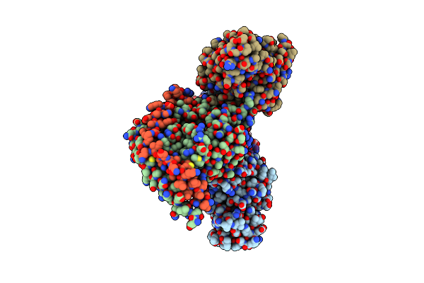
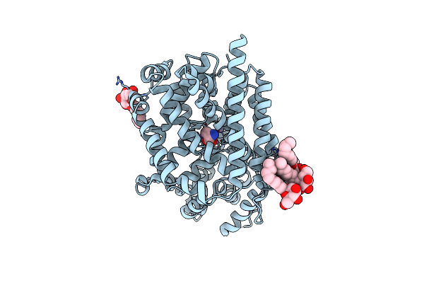
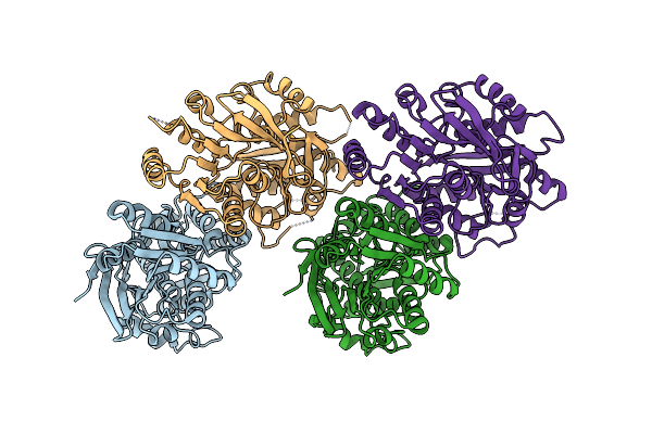
Organism: Homo sapiens
Method: X-RAY DIFFRACTION Resolution:2.85 Å Release Date: 2025-02-26 Classification: HORMONE Ligands: IPH, ZN, CL, MYR |
 |
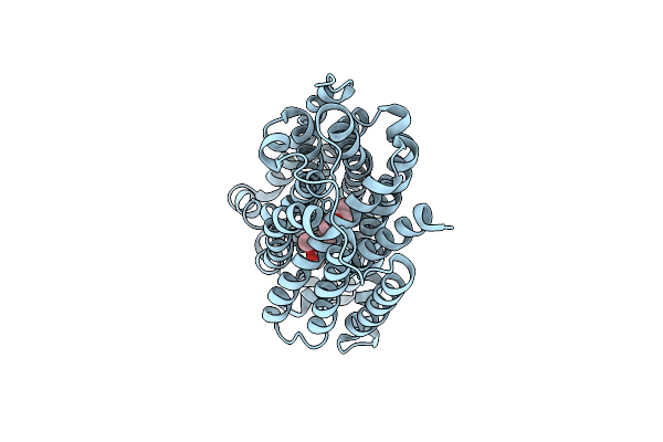
Organism: Homo sapiens, Mus musculus
Method: ELECTRON MICROSCOPY Release Date: 2023-11-01 Classification: SIGNALING PROTEIN Ligands: CLR, PLM |
 |
Structure Of The Human Vesicular Monoamine Transporter 2 (Vmat2) Bound To Tetrabenazine In An Occluded Conformation
Organism: Aequorea victoria, Homo sapiens, Vicugna pacos
Method: ELECTRON MICROSCOPY Release Date: 2023-10-25 Classification: TRANSPORT PROTEIN Ligands: EBZ |
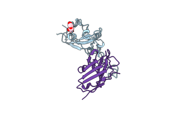 |
Cryo-Em Structure Of Sars-Cov-2 Beta (B.1.351) Spike Protein In Complex With Vh Domain F6 (Focused Refinement Of Rbd And Vh F6)
Organism: Severe acute respiratory syndrome coronavirus 2, Homo sapiens
Method: ELECTRON MICROSCOPY Release Date: 2022-08-24 Classification: VIRAL PROTEIN/IMMUNE SYSTEM Ligands: NAG |
 |
Organism: Aquifex aeolicus (strain vf5)
Method: X-RAY DIFFRACTION Resolution:2.14 Å Release Date: 2021-05-19 Classification: TRANSPORT PROTEIN Ligands: ALA, NA, BOG |
 |
Organism: Aquifex aeolicus (strain vf5)
Method: X-RAY DIFFRACTION Resolution:2.10 Å Release Date: 2021-05-19 Classification: TRANSPORT PROTEIN Ligands: ALA, NA, BOG |
 |
Organism: Aquifex aeolicus (strain vf5)
Method: X-RAY DIFFRACTION Resolution:2.60 Å Release Date: 2021-05-19 Classification: TRANSPORT PROTEIN |
 |
Organism: Rattus norvegicus
Method: X-RAY DIFFRACTION Resolution:2.51 Å Release Date: 2018-12-19 Classification: MEMBRANE PROTEIN Ligands: NAG |
 |
Organism: Rattus norvegicus
Method: X-RAY DIFFRACTION Resolution:1.96 Å Release Date: 2018-12-19 Classification: MEMBRANE PROTEIN Ligands: NAG, PO4, DMS, GOL |
 |
Organism: Rattus norvegicus
Method: X-RAY DIFFRACTION Resolution:2.25 Å Release Date: 2012-10-24 Classification: TRANSPORT PROTEIN Ligands: NAG |
 |
Organism: Rattus norvegicus
Method: X-RAY DIFFRACTION Resolution:2.20 Å Release Date: 2011-03-09 Classification: TRANSPORT PROTEIN Ligands: NAG, PO4 |
 |
Organism: Rattus norvegicus
Method: X-RAY DIFFRACTION Resolution:4.20 Å Release Date: 2011-03-09 Classification: TRANSPORT PROTEIN |