
Search Count: 13
 |
Organism: Methanocaldococcus jannaschii
Method: X-RAY DIFFRACTION Resolution:2.00 Å Release Date: 2008-03-18 Classification: LIGASE Ligands: BTN |
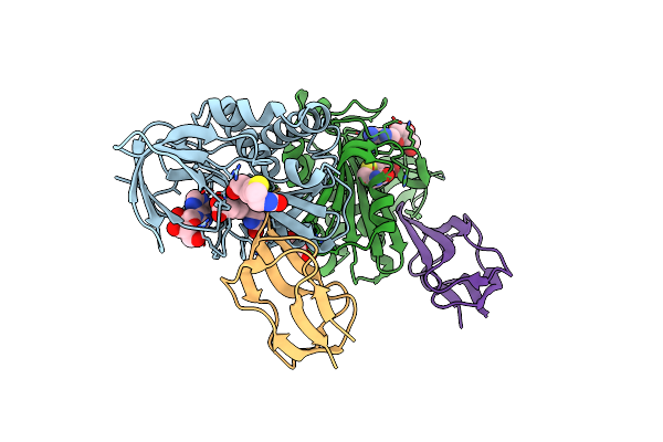 |
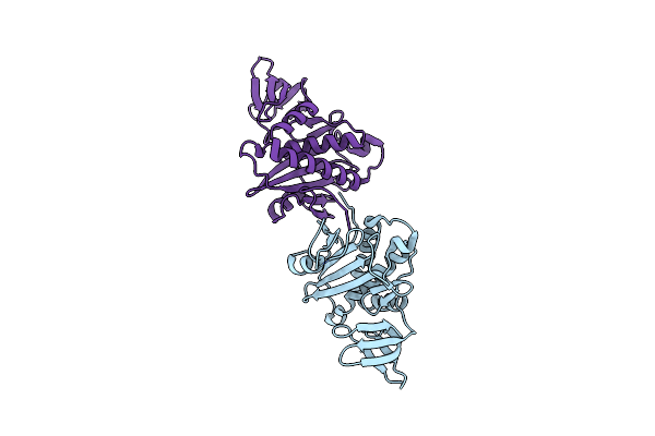
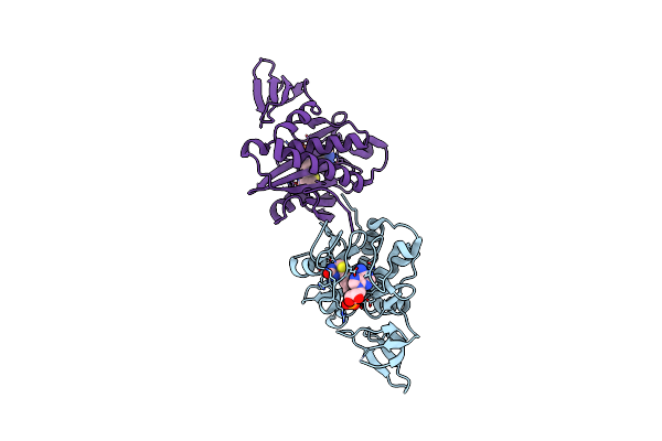
Crystal Structure Of The Biotin Protein Ligase (Mutations R48A And K111A) And Biotin Carboxyl Carrier Protein Complex From Pyrococcus Horikoshii Ot3
Organism: Pyrococcus horikoshii
Method: X-RAY DIFFRACTION Resolution:2.00 Å Release Date: 2008-03-18 Classification: LIGASE Ligands: BTN, ADN, GOL |
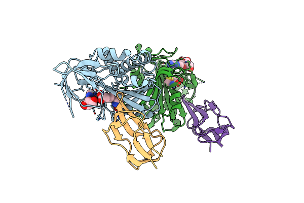 |
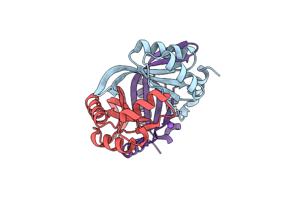
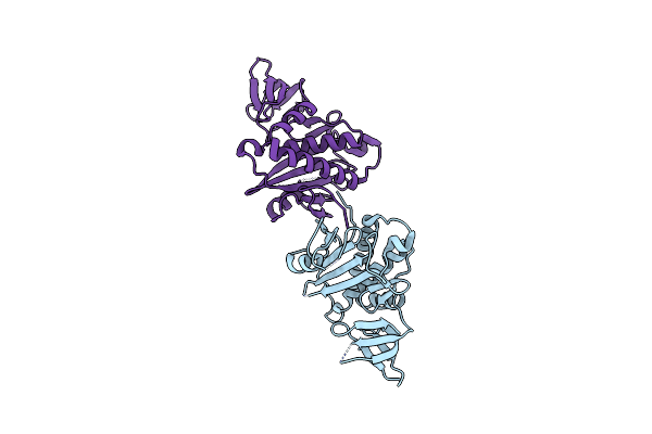
Crystal Structure Of The Biotin Protein Ligase (Mutation R48A) And Biotin Carboxyl Carrier Protein Complex From Pyrococcus Horikoshii Ot3
Organism: Pyrococcus horikoshii
Method: X-RAY DIFFRACTION Resolution:2.71 Å Release Date: 2008-03-18 Classification: LIGASE Ligands: BTN, ADN |
 |
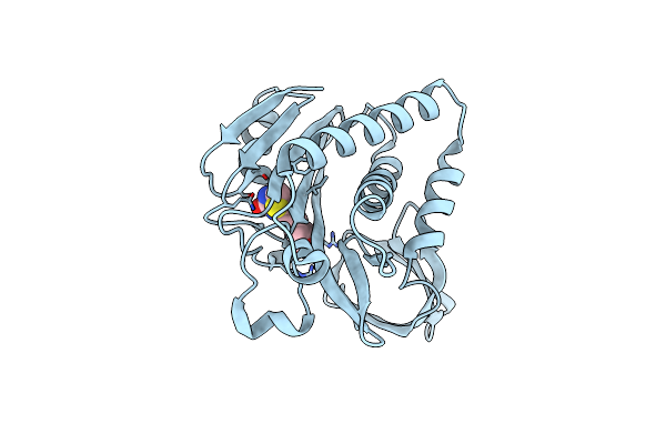
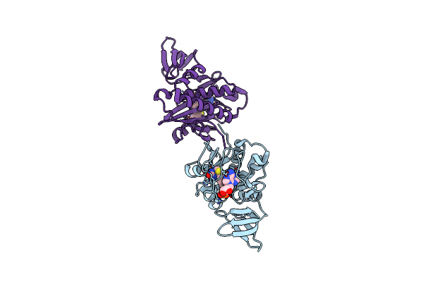
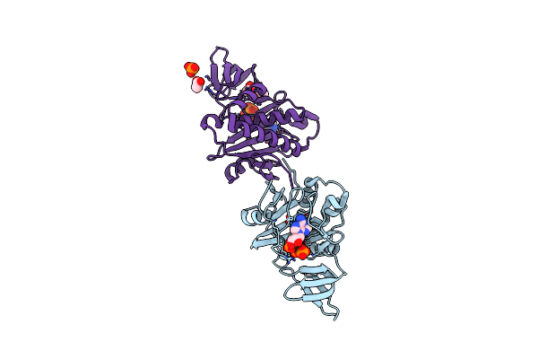
Crystal Structure Of Biotin Protein Ligase From Pyrococcus Horikoshii Complexed With Adenosine And Biotin, Mutations R48A And K111A
Organism: Pyrococcus horikoshii
Method: X-RAY DIFFRACTION Resolution:1.50 Å Release Date: 2008-02-05 Classification: LIGASE Ligands: ADN, BTN |
 |
Organism: Homo sapiens
Method: X-RAY DIFFRACTION Resolution:2.05 Å Release Date: 2008-01-22 Classification: STRUCTURAL GENOMICS, UNKNOWN FUNCTION |
 |
Crystal Structure Of Biotin Protein Ligase From Pyrococcus Horikoshii, Mutations R48A And K111A
Organism: Pyrococcus horikoshii
Method: X-RAY DIFFRACTION Resolution:1.50 Å Release Date: 2007-06-26 Classification: LIGASE |
 |
Organism: Pyrococcus horikoshii
Method: X-RAY DIFFRACTION Resolution:2.00 Å Release Date: 2007-06-26 Classification: STRUCTURAL GENOMICS, UNKNOWN FUNCTION |
 |
Crystal Structure Of Biotin Protein Ligase From Pyrococcus Horikoshii Complexed With The Reaction Product Analog Biotinol-5'-Amp, Mutations R48A And K111A
Organism: Pyrococcus horikoshii
Method: X-RAY DIFFRACTION Resolution:1.75 Å Release Date: 2007-06-05 Classification: LIGASE Ligands: BTX |
 |
Crystal Structure Of Biotin Protein Ligase From Pyrococcus Horikoshii, Mutation R48A
Organism: Pyrococcus horikoshii
Method: X-RAY DIFFRACTION Resolution:1.45 Å Release Date: 2007-03-27 Classification: LIGASE |
 |
Crystal Structure Of Biotin Protein Ligase From Pyrococcus Horikoshii Complexed With Biotinyl-5'-Amp, Mutation R48A
Organism: Pyrococcus horikoshii
Method: X-RAY DIFFRACTION Resolution:1.28 Å Release Date: 2007-03-01 Classification: LIGASE Ligands: BT5 |
 |
Crystal Structure Of Biotin Protein Ligase From Pyrococcus Horikoshii Ot3 In Complex With Atp
Organism: Pyrococcus horikoshii
Method: X-RAY DIFFRACTION Resolution:2.00 Å Release Date: 2006-05-23 Classification: LIGASE Ligands: ATP, PO4, ACY |
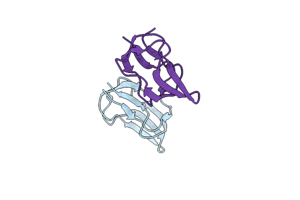 |
Structure Of Biotin Carboxyl Carrier Protein (74Val Start) From Pyrococcus Horikoshi Ot3 Ligand Free Form Ii
Organism: Pyrococcus horikoshii
Method: X-RAY DIFFRACTION Resolution:1.55 Å Release Date: 2006-05-01 Classification: LIPID BINDING PROTEIN,TRANSFERASE |
 |
Structure Of Biotin Carboxyl Carrier Protein (74Val Start) From Pyrococcus Horikoshi Ot3 Ligand Free Form I
Organism: Pyrococcus horikoshii
Method: X-RAY DIFFRACTION Resolution:1.55 Å Release Date: 2006-05-01 Classification: LIPID BINDING PROTEIN,TRANSFERASE |