
Search Count: 8
 |
Organism: Streptomyces rubiginosus
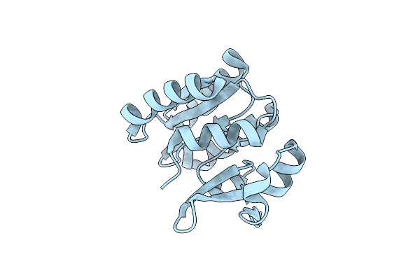
Method: X-RAY DIFFRACTION Resolution:1.75 Å Release Date: 2018-11-28 Classification: ISOMERASE Ligands: ACT, MN, EDO |
 |
Organism: Streptomyces rubiginosus
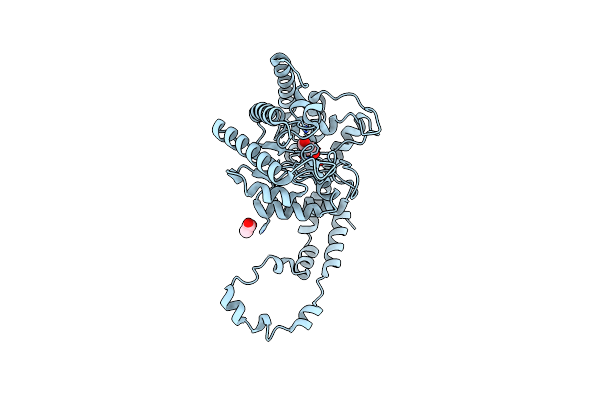
Method: X-RAY DIFFRACTION Resolution:1.40 Å Release Date: 2018-11-28 Classification: ISOMERASE Ligands: MG, ACT |
 |
Organism: Streptomyces rubiginosus
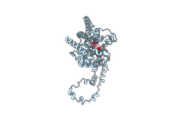
Method: X-RAY DIFFRACTION Resolution:1.40 Å Release Date: 2018-11-28 Classification: ISOMERASE Ligands: MN, GLC |
 |
Organism: Rhodothermus marinus (strain atcc 43812 / dsm 4252 / r-10)
Method: X-RAY DIFFRACTION Resolution:1.87 Å Release Date: 2018-05-30 Classification: PROTEIN TRANSPORT |
 |
Crystal Structure Of Glucose Isomerase In Complex With Glycerol In One Metal Binding Mode
Organism: Streptomyces rubiginosus
Method: X-RAY DIFFRACTION Resolution:1.91 Å Release Date: 2017-09-20 Classification: ISOMERASE Ligands: ACT, MG, GOL |
 |
Crystal Structure Of Glucose Isomerase In Complex With Xylitol Inhibitor In One Metal Binding Mode
Organism: Streptomyces rubiginosus
Method: X-RAY DIFFRACTION Resolution:1.40 Å Release Date: 2017-09-20 Classification: ISOMERASE Ligands: MG, XYL |
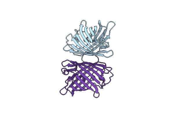 |
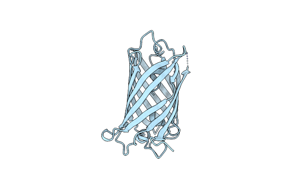
Organism: Zoanthus sp.
Method: X-RAY DIFFRACTION Resolution:2.90 Å Release Date: 2017-09-13 Classification: FLUORESCENT PROTEIN |
 |
Organism: Zoanthus sp.
Method: X-RAY DIFFRACTION Resolution:2.30 Å Release Date: 2017-09-13 Classification: FLUORESCENT PROTEIN |