
Search Count: 13
 |
Organism: Drosophila melanogaster
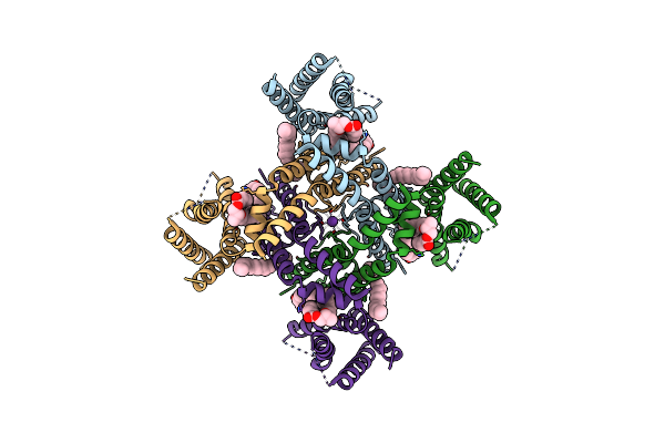
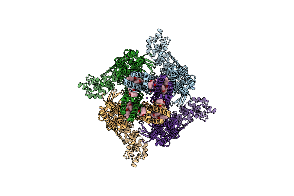
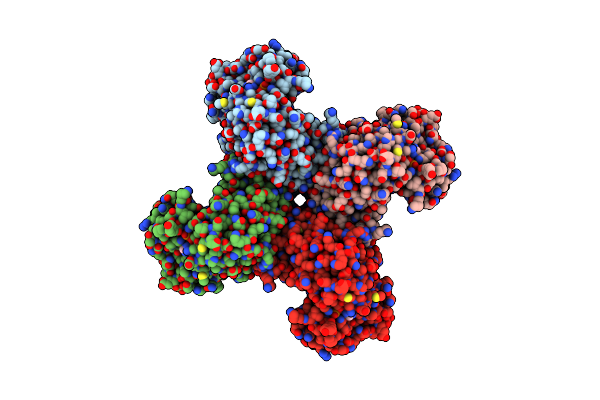
Method: ELECTRON MICROSCOPY Release Date: 2023-12-20 Classification: MEMBRANE PROTEIN Ligands: POV, K |
 |
Organism: Rattus norvegicus
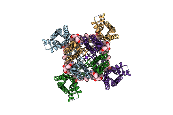
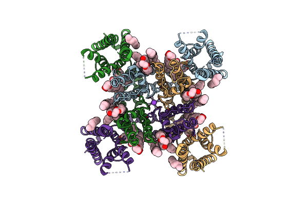
Method: ELECTRON MICROSCOPY Release Date: 2023-09-27 Classification: MEMBRANE PROTEIN Ligands: POV, K |
 |
Organism: Rattus norvegicus
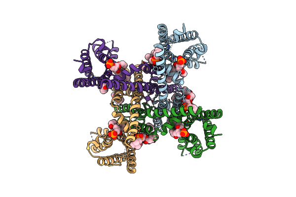
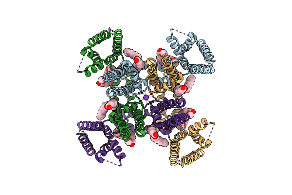
Method: ELECTRON MICROSCOPY Release Date: 2023-09-27 Classification: MEMBRANE PROTEIN Ligands: POV, K |
 |
Organism: Rattus norvegicus
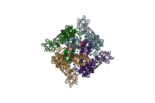
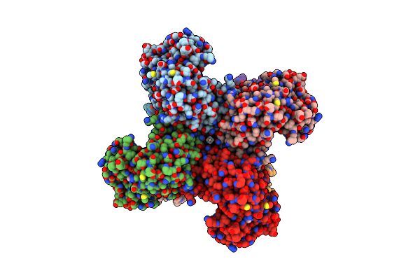
Method: ELECTRON MICROSCOPY Release Date: 2023-05-31 Classification: MEMBRANE PROTEIN Ligands: P0T, NA |
 |
Organism: Rattus norvegicus
Method: ELECTRON MICROSCOPY Release Date: 2023-05-31 Classification: MEMBRANE PROTEIN Ligands: P0T, NA, POV |
 |
Organism: Drosophila melanogaster
Method: ELECTRON MICROSCOPY Release Date: 2022-03-30 Classification: MEMBRANE PROTEIN Ligands: POV, K |
 |
Organism: Drosophila melanogaster
Method: ELECTRON MICROSCOPY Release Date: 2022-03-30 Classification: MEMBRANE PROTEIN Ligands: POV, K |
 |
Organism: Rattus norvegicus
Method: ELECTRON MICROSCOPY Release Date: 2018-08-22 Classification: MEMBRANE PROTEIN Ligands: NAP |
 |
The Voltage-Activated Kv1.2-2.1 Paddle Chimera Channel In Lipid Nanodiscs, Cytosolic Domain
Organism: Rattus norvegicus
Method: ELECTRON MICROSCOPY Release Date: 2018-08-22 Classification: MEMBRANE PROTEIN Ligands: NAP |
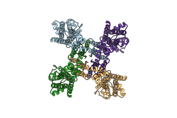 |
The Voltage-Activated Kv1.2-2.1 Paddle Chimera Channel In Lipid Nanodiscs, Transmembrane Domain Of Subunit Alpha
Organism: Rattus norvegicus
Method: ELECTRON MICROSCOPY Release Date: 2018-08-22 Classification: MEMBRANE PROTEIN |
 |
 |
 |
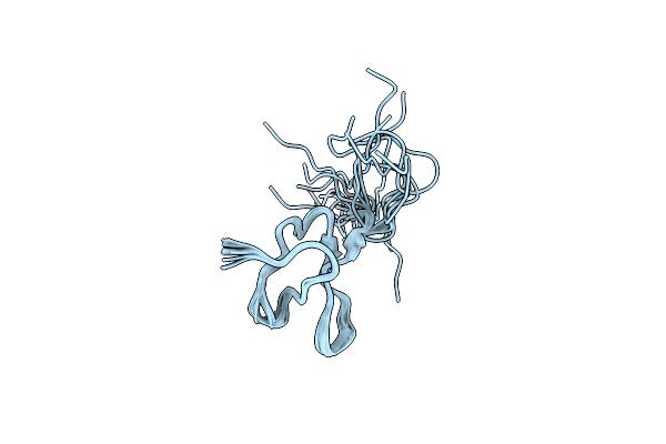
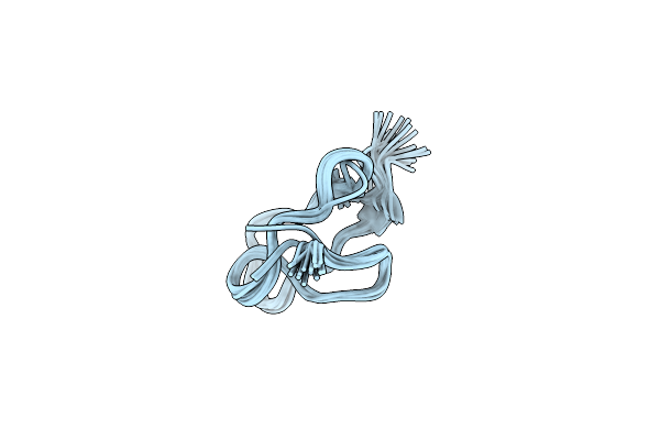
Organism: Plesiophrictus guangxiensis
Method: SOLUTION NMR Release Date: 2010-05-26 Classification: MEMBRANE PROTEIN INHIBITOR |