
Search Count: 16
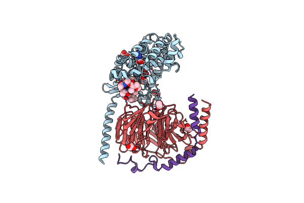 |
Crystal Structure Of The G11 Protein Heterotrimer Bound To Ym-254890 Inhibitor
Organism: Homo sapiens
Method: X-RAY DIFFRACTION Resolution:1.70 Å Release Date: 2025-03-19 Classification: SIGNALING PROTEIN Ligands: GDP, EDO, DAM, HF2, THC, OTH, HL2, MAA, ALA, ACE |
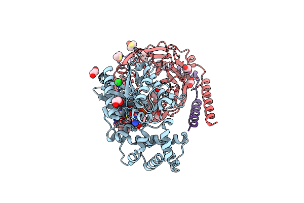 |
Crystal Structure Of The G11 Protein Heterotrimer Bound To Fr900359 Inhibitor
Organism: Homo sapiens
Method: X-RAY DIFFRACTION Resolution:1.43 Å Release Date: 2025-03-19 Classification: SIGNALING PROTEIN Ligands: EDO, GDP, ZN, CL, DAM, HF2, UDL, OTH, HL2, MAA, ALA, PPI, DMS |
 |
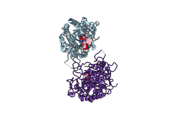
Dark Structure Of The Human Adenosine A2A Receptor Bound To Synthetic Photoswitch "Stilswitch3" Determined By Serial Synchrotron Crystallography
Organism: Homo sapiens
Method: X-RAY DIFFRACTION Resolution:2.65 Å Release Date: 2025-01-08 Classification: MEMBRANE PROTEIN Ligands: OLA, A1H3H, CLR, NA |
 |
Crystal Structure Of The Adenosine A2A Receptor In Complex With Istradefylline
Organism: Homo sapiens
Method: X-RAY DIFFRACTION Resolution:1.94 Å Release Date: 2025-01-08 Classification: MEMBRANE PROTEIN Ligands: JQ9, CLR, OLA, NA |
 |
Crystal Structure Of The Adenosine A2A Receptor In Complex With The Synthetic Photoswitch 'Azoswitch2
Organism: Homo sapiens
Method: X-RAY DIFFRACTION Resolution:2.20 Å Release Date: 2025-01-08 Classification: MEMBRANE PROTEIN Ligands: A1H3L, CLR, OLA, NA |
 |
Crystal Structure Of The Adenosine A2A Receptor In Complex With The Synthetic Photoswitch 'Stilswitch1
Organism: Homo sapiens
Method: X-RAY DIFFRACTION Resolution:2.25 Å Release Date: 2025-01-08 Classification: MEMBRANE PROTEIN Ligands: A1H3J, CLR, OLA, OLC, OLB, NA |
 |
Crystal Structure Of The Adenosine A2A Receptor In Complex With The Synthetic Photoswitch 'Stilswitch2
Organism: Homo sapiens
Method: X-RAY DIFFRACTION Resolution:2.31 Å Release Date: 2025-01-08 Classification: MEMBRANE PROTEIN Ligands: A1H3I, CLR, OLC, OLA, NA |
 |
Crystal Structure Of The Adenosine A2A Receptor In Complex With The Synthetic Photoswitch 'Stilswitch3
Organism: Homo sapiens
Method: X-RAY DIFFRACTION Resolution:2.05 Å Release Date: 2025-01-08 Classification: MEMBRANE PROTEIN Ligands: A1H3H, CLR, OLA, NA |
 |
Crystal Structure Of The Adenosine A2A Receptor In Complex With The Synthetic Photoswitch 'Stilswitch4
Organism: Homo sapiens
Method: X-RAY DIFFRACTION Resolution:2.20 Å Release Date: 2025-01-08 Classification: MEMBRANE PROTEIN Ligands: A1H3K, CLR, OLA, OLC, NA |
 |
Dark Structure Of The Human Adenosine A2A Receptor Bound To Synthetic Photoswitch 'Stilswitch2' Determined By Serial Synchrotron Crystallography
Organism: Homo sapiens
Method: X-RAY DIFFRACTION Resolution:2.45 Å Release Date: 2025-01-08 Classification: MEMBRANE PROTEIN Ligands: A1H3I, CLR, OLA, OLB, OLC, NA |
 |
Steady State Structure Of The Human Adenosine A2A Receptor Bound To Synthetic Photoswitch 'Stilswitch2' Determined By Serial Synchrotron Crystallography
Organism: Homo sapiens
Method: X-RAY DIFFRACTION Resolution:2.80 Å Release Date: 2025-01-08 Classification: MEMBRANE PROTEIN Ligands: A1H3I, CLR, OLA, NA |
 |
Steady State Structure Of The Human Adenosine A2A Receptor Bound To Synthetic Photoswitch 'Stilswitch3' Determined By Serial Synchrotron Crystallography
Organism: Homo sapiens, Escherichia coli
Method: X-RAY DIFFRACTION Resolution:3.05 Å Release Date: 2025-01-08 Classification: MEMBRANE PROTEIN Ligands: A1H3H, OLA, NA |
 |
Crystal Structure Of Ene-Reductase Oye4 From Botryotinia Fuckeliana (Bfoye4)
Organism: Botryotinia fuckeliana (strain b05.10)
Method: X-RAY DIFFRACTION Resolution:2.15 Å Release Date: 2022-07-13 Classification: OXIDOREDUCTASE Ligands: FMN, FMT |
 |
Crystal Structure Of G. Sulphuraria Ene-Reductase Gsoye In Complex With B-Angelica Lactone
Organism: Galdieria sulphuraria
Method: X-RAY DIFFRACTION Resolution:1.50 Å Release Date: 2022-07-13 Classification: OXIDOREDUCTASE Ligands: U5N, FMN |
 |
Organism: Saccharomyces cerevisiae
Method: X-RAY DIFFRACTION Resolution:2.45 Å Release Date: 2022-07-13 Classification: OXIDOREDUCTASE Ligands: FMN, GOL |
 |
Crystal Structure Of Ene-Reductase Gsoye From Galdieria Sulphuraria In Complex With Alpha-Angelica Lactone
Organism: Galdieria sulphuraria
Method: X-RAY DIFFRACTION Resolution:1.63 Å Release Date: 2022-07-13 Classification: OXIDOREDUCTASE Ligands: U6W, FMN |