
Search Count: 51
 |
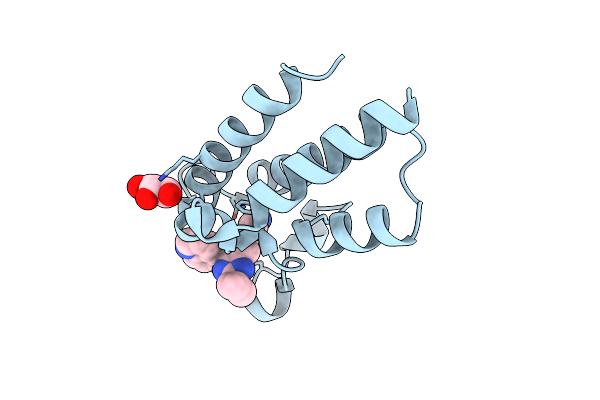
Organism: Homo sapiens
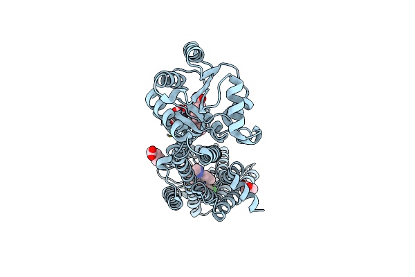
Method: X-RAY DIFFRACTION Resolution:1.91 Å Release Date: 2024-09-04 Classification: ANTIBIOTIC Ligands: MYR, A1AZR |
 |
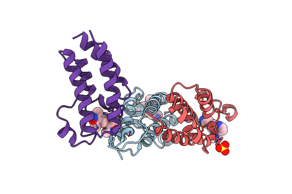
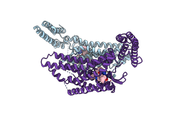
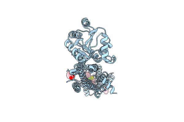
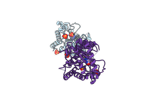
Organism: Acinetobacter baumannii
Method: X-RAY DIFFRACTION Resolution:2.80 Å Release Date: 2024-09-04 Classification: ANTIBIOTIC Ligands: ANP, MVC, MG, A1AZS |
 |
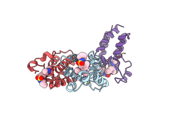
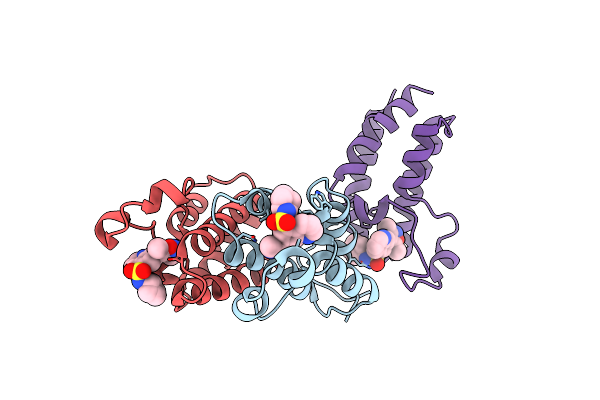
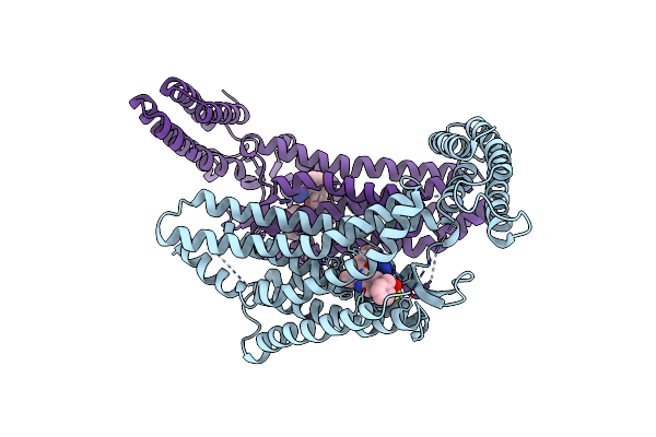
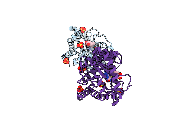
Organism: Acinetobacter baumannii
Method: ELECTRON MICROSCOPY Release Date: 2024-04-24 Classification: ANTIMICROBIAL PROTEIN Ligands: ANP, ZQF |
 |
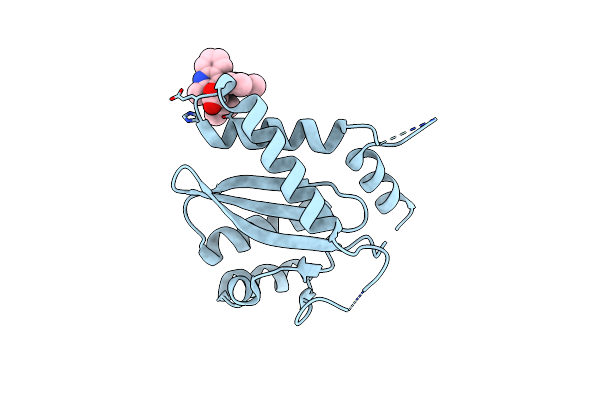
N-Terminal Bromodomain Of Human Brd2 With N-((4-(3-(N-Cyclopentylsulfamoyl)-4-Methylphenyl)-3-Methylisoxazol-5-Yl)Methyl)Acetamide Inhibitor
Organism: Homo sapiens
Method: X-RAY DIFFRACTION Resolution:1.85 Å Release Date: 2019-01-23 Classification: transcription/transcription inhibitor Ligands: JWA |
 |
N-Terminal Bromodomain Of Human Brd2 In Complex With 4,4'-(Quinoline-5,7-Diyl)Bis(3,5-Dimethylisoxazole) Inhibitor
Organism: Homo sapiens
Method: X-RAY DIFFRACTION Resolution:1.80 Å Release Date: 2019-01-23 Classification: transcription/transcription inhibitor Ligands: JWD, SO4 |
 |
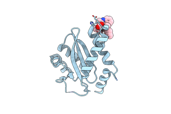
N-Terminal Bromodomain Of Human Brd2 In Complex With N-Cyclopentyl-7-(3,5-Dimethylisoxazol-4-Yl)Quinoline-5-Sulfonamide Inhibitor
Organism: Homo sapiens
Method: X-RAY DIFFRACTION Resolution:1.80 Å Release Date: 2019-01-23 Classification: transcription/transcription inhibitor Ligands: JVY |
 |
C-Terminal Bromodomain Of Human Brd2 In Complex With 4-(2-Cyclopropyl-7-(6-Methylquinolin-5-Yl)-1H-Benzo[D]Imidazol-5-Yl)-3,5-Dimethylisoxazole Inhibitor
Organism: Homo sapiens
Method: X-RAY DIFFRACTION Resolution:1.27 Å Release Date: 2019-01-23 Classification: transcription/transcription inhibitor Ligands: JW4, GOL |
 |
Xfel Structure Of Human Angiotensin Ii Type 2 Receptor (Monoclinic Form) In Complex With Compound 1 (N-Benzyl-N-(2-Ethyl-4-Oxo-3-{[2'-(2H-Tetrazol-5-Yl)[1,1'-Biphenyl]-4-Yl])
Organism: Escherichia coli, Homo sapiens
Method: X-RAY DIFFRACTION Resolution:2.80 Å Release Date: 2017-04-05 Classification: SIGNALING PROTEIN Ligands: 8ES |
 |
Xfel Structure Of Human Angiotensin Ii Type 2 Receptor (Orthorhombic Form) In Complex With Compound 1 (N-Benzyl-N-(2-Ethyl-4-Oxo-3-{[2'-(2H-Tetrazol-5-Yl)[1,1'-Biphenyl]-4-Yl] Methyl}-3,4-Dihydroquinazolin-6-Yl)Thiophene-2-Carboxamide)
Organism: Escherichia coli, Homo sapiens
Method: X-RAY DIFFRACTION Resolution:2.80 Å Release Date: 2017-04-05 Classification: SIGNALING PROTEIN Ligands: 8ES, OLC, OLA |
 |
Synchrotron Structure Of Human Angiotensin Ii Type 2 Receptor In Complex With Compound 2 (N-[(Furan-2-Yl)Methyl]-N-(4-Oxo-2-Propyl-3-{[2'-(2H-Tetrazol-5-Yl)[1,1'- Biphenyl]-4-Yl]Methyl}-3,4-Dihydroquinazolin-6-Yl)Benzamide)
Organism: Escherichia coli, Homo sapiens
Method: X-RAY DIFFRACTION Resolution:2.90 Å Release Date: 2017-04-05 Classification: SIGNALING PROTEIN Ligands: 8EM |
 |
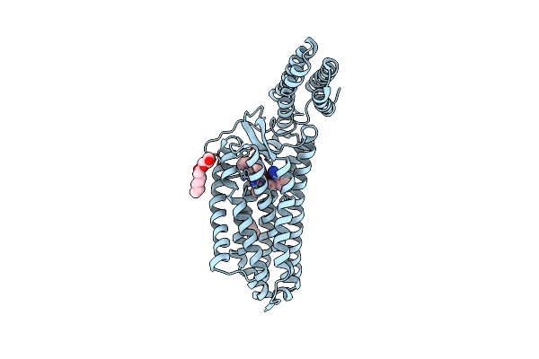
Structures Of The Human Ox1 Orexin Receptor Bound To Selective And Dual Antagonists
Organism: Homo sapiens, Pyrococcus abyssi (strain ge5 / orsay)
Method: X-RAY DIFFRACTION Resolution:2.75 Å Release Date: 2016-03-09 Classification: SIGNALING PROTEIN Ligands: SUV, OLA |
 |
Structures Of The Human Ox1 Orexin Receptor Bound To Selective And Dual Antagonists
Organism: Homo sapiens, Pyrococcus abyssi (strain ge5 / orsay)
Method: X-RAY DIFFRACTION Resolution:2.83 Å Release Date: 2016-03-09 Classification: SIGNALING PROTEIN Ligands: 4OT, OLA |
 |
Organism: Human immunodeficiency virus type 1
Method: X-RAY DIFFRACTION Resolution:1.90 Å Release Date: 2012-04-25 Classification: TRANSFERASE/TRANSFERASE INHIBITOR Ligands: TQ2 |
 |
Organism: Human immunodeficiency virus type 1
Method: X-RAY DIFFRACTION Resolution:2.00 Å Release Date: 2012-04-25 Classification: TRANSFERASE/TRANSFERASE INHIBITOR Ligands: TQX |
 |
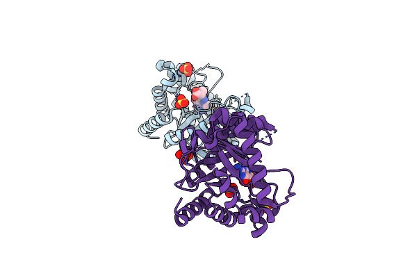
Crystal Structure Of Ampc Beta-Lactamase In Complex With A Sulfonamide Boronic Acid Inhibitor
Organism: Escherichia coli
Method: X-RAY DIFFRACTION Resolution:1.60 Å Release Date: 2010-11-03 Classification: HYDROLASE Ligands: BSF, PO4 |
 |
Crystal Structure Of Ampc Beta-Lactamase In Complex With A Sulfonamide Boronic Acid Inhibitor
Organism: Escherichia coli
Method: X-RAY DIFFRACTION Resolution:1.78 Å Release Date: 2010-11-03 Classification: HYDROLASE Ligands: BSG, PO4 |
 |
Crystal Structure Of Ampc Beta-Lactamase In Complex With A Sulfonamide Boronic Acid Inhibitor
Organism: Escherichia coli
Method: X-RAY DIFFRACTION Resolution:1.64 Å Release Date: 2010-11-03 Classification: HYDROLASE Ligands: BSH, PO4 |
 |
Organism: Bacillus anthracis str. a2012
Method: X-RAY DIFFRACTION Resolution:2.32 Å Release Date: 2009-12-08 Classification: Transferase/Transferase Inhibitor Ligands: B52, SO4 |
 |
Organism: Bacillus anthracis str. a2012
Method: X-RAY DIFFRACTION Resolution:2.40 Å Release Date: 2009-12-08 Classification: Transferase/Transferase Inhibitor Ligands: B53, SO4 |
 |
Organism: Bacillus anthracis str. a2012
Method: X-RAY DIFFRACTION Resolution:2.20 Å Release Date: 2009-12-08 Classification: Transferase/Transferase Inhibitor Ligands: B54, SO4 |