
Search Count: 17
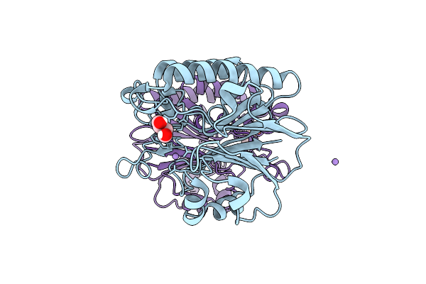 |
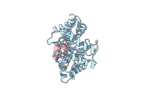
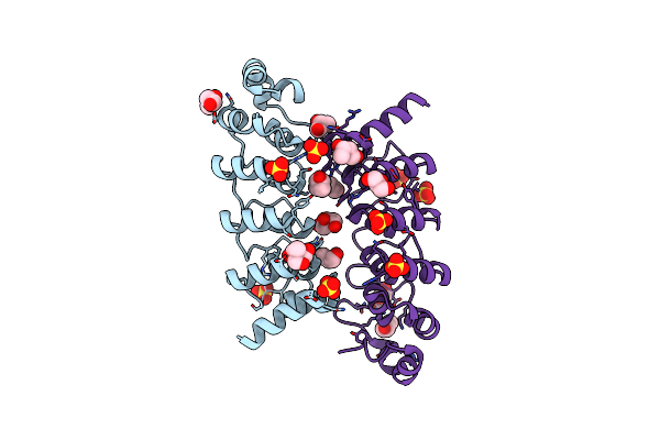
Crystal Structure Of Hp1526 (Xtha)- A Base Excision Dna Repair Protein In Helicobacter Pylori
Organism: Helicobacter pylori 26695
Method: X-RAY DIFFRACTION Resolution:1.84 Å Release Date: 2023-12-27 Classification: STRUCTURAL PROTEIN Ligands: MN, BU2 |
 |
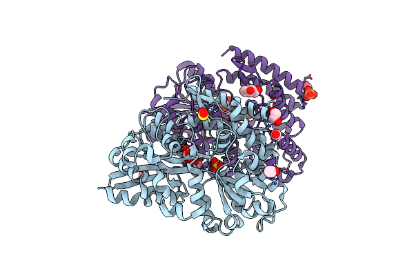
Organism: Streptomyces cattleya
Method: X-RAY DIFFRACTION Resolution:1.70 Å Release Date: 2022-04-13 Classification: BIOSYNTHETIC PROTEIN Ligands: GOL, ACT, NA, MG, BU2 |
 |
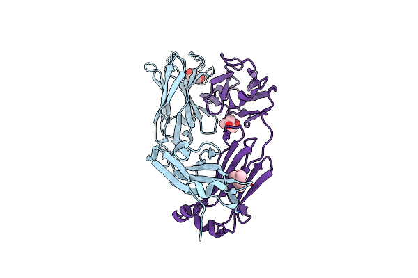
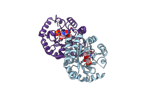
Structure Of Sybody Mr17-Sr31 Fusion In Complex With The Sars-Cov-2 S Receptor Binding Domain (Rbd)
Organism: Synthetic construct, Severe acute respiratory syndrome coronavirus 2
Method: X-RAY DIFFRACTION Resolution:2.10 Å Release Date: 2021-02-17 Classification: PROTEIN BINDING Ligands: GOL, ACT, 1PE, BU2, CD |
 |
Crystal Structure Of Beta-Glycosides-Binding Protein (W177X) Of Abc Transporter In An Open-Liganded State Bound To Sophorose
Organism: Thermus thermophilus (strain hb8 / atcc 27634 / dsm 579)
Method: X-RAY DIFFRACTION Resolution:1.70 Å Release Date: 2020-09-16 Classification: SUGAR BINDING PROTEIN Ligands: SO2, CO2, ACT, SO4, EDO, BU2 |
 |
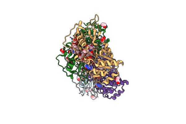
Organism: Homo sapiens
Method: X-RAY DIFFRACTION Resolution:2.70 Å Release Date: 2020-02-12 Classification: IMMUNE SYSTEM Ligands: BU2, MPD |
 |
Human Sirt5 In Complex With Stalled Peptidylimidate Intermediate Of Inhibitory Compound 29
Organism: Homo sapiens
Method: X-RAY DIFFRACTION Resolution:1.32 Å Release Date: 2017-11-01 Classification: SIGNALING PROTEIN Ligands: ZN, BV8, EDO, BU2, DMS |
 |
Sulphotransferase-18 From Arabidopsis Thaliana In Complex With 3'-Phosphoadenosine 5'-Phosphate (Pap)And Sinigrin
Organism: Arabidopsis thaliana
Method: X-RAY DIFFRACTION Resolution:1.92 Å Release Date: 2017-07-05 Classification: TRANSFERASE Ligands: PAP, SZZ, EDO, BU2 |
 |
Native Structure Of The Linalool Dehydratase-Isomerase From Castellaniella Defragrans
Organism: Castellaniella defragrans
Method: X-RAY DIFFRACTION Resolution:2.10 Å Release Date: 2016-06-01 Classification: LYASE, ISOMERASE Ligands: BU2, CL, K |
 |
Crystal Structure Of Deoxyribose-Phosphate Aldolase From Escherichia Coli (K58E-Y96W Mutant)
Organism: Escherichia coli
Method: X-RAY DIFFRACTION Resolution:1.10 Å Release Date: 2016-05-04 Classification: LYASE Ligands: BU2, EPE |
 |
Crystal Structure Of Legionella Pneumophila Dephospho-Coa Kinase In Complex With Bu2
Organism: Legionella pneumophila subsp. pneumophila
Method: X-RAY DIFFRACTION Resolution:2.10 Å Release Date: 2014-12-10 Classification: TRANSFERASE Ligands: BU2, PO4 |
 |
Organism: Homo sapiens, Synthetic construct
Method: X-RAY DIFFRACTION Resolution:2.42 Å Release Date: 2014-10-08 Classification: TRANSFERASE/TRANSFERASE INHIBITOR Ligands: BU2, GOL |
 |
Disulfide Isomerase From Multidrug Resistance Inca/C Related Integrative And Conjugative Elements In Oxidized State (P21 Space Group)
Organism: Proteus mirabilis
Method: X-RAY DIFFRACTION Resolution:2.21 Å Release Date: 2013-12-11 Classification: ISOMERASE Ligands: BU2 |
 |
Crystal Structure Of Sulfide:Quinone Oxidoreductase From Acidithiobacillus Ferrooxidans
Organism: Acidithiobacillus ferrooxidans
Method: X-RAY DIFFRACTION Resolution:2.30 Å Release Date: 2011-08-17 Classification: OXIDOREDUCTASE Ligands: FAD, BU2, SO4, H2S, S3H |
 |
Organism: Arabidopsis thaliana
Method: X-RAY DIFFRACTION Resolution:1.85 Å Release Date: 2009-12-08 Classification: SIGNALING PROTEIN Ligands: BU2 |
 |
Crystal Structure Of The Dimethylallyl Tryptophan Synthase Fgapt2 From Aspergillus Fumigatus
Organism: Aspergillus fumigatus
Method: X-RAY DIFFRACTION Resolution:1.76 Å Release Date: 2009-09-01 Classification: TRANSFERASE Ligands: BU2, GOL |
 |
Organism: Thermoplasma volcanium
Method: X-RAY DIFFRACTION Resolution:1.65 Å Release Date: 2008-03-11 Classification: PROTEIN BINDING Ligands: SO4, 144, BU2, GOL, CL |
 |
Crystal Structure Of Orotidine Monophosphate Decarboxylase Complex With Xmp
Organism: Methanothermobacter thermautotrophicus str. delta h
Method: X-RAY DIFFRACTION Resolution:1.90 Å Release Date: 2002-08-07 Classification: LYASE Ligands: BU2, XMP |