
Search Count: 87
 |
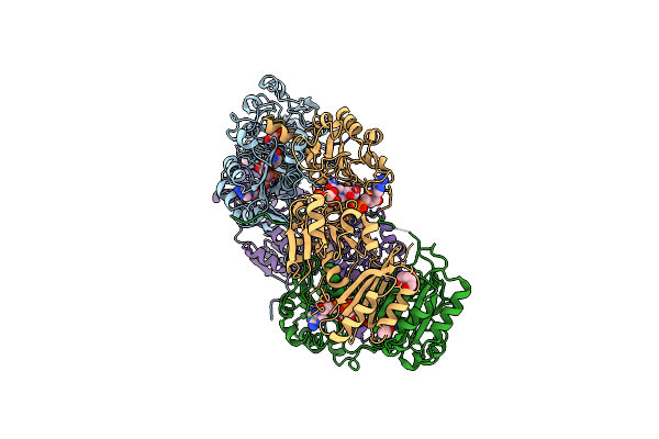
Crystal Structure Of Bf3526 Peptidase From Bacteroides Fragilis In Complex With A Peptide
Organism: Bacteroides fragilis nctc 9343, Synthetic construct
Method: X-RAY DIFFRACTION Release Date: 2025-07-16 Classification: HYDROLASE Ligands: K, ZN, EDO, POL, PEG, BU1, HEZ |
 |
Organism: Oryza sativa japonica group
Method: X-RAY DIFFRACTION Release Date: 2025-05-21 Classification: HYDROLASE Ligands: A1EBP, GOL, BEZ, PDO, BU1, PGO |
 |
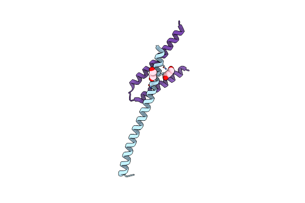
Organism: Homo sapiens, Drosophila melanogaster
Method: X-RAY DIFFRACTION Resolution:2.64 Å Release Date: 2025-01-29 Classification: RNA BINDING PROTEIN Ligands: BU1 |
 |
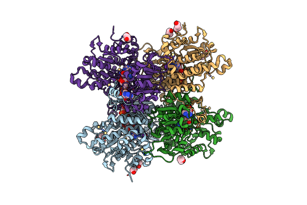
Organism: Saccharomyces cerevisiae
Method: X-RAY DIFFRACTION Resolution:2.70 Å Release Date: 2025-01-15 Classification: CYTOSOLIC PROTEIN Ligands: NAD, 3PG, BU1 |
 |
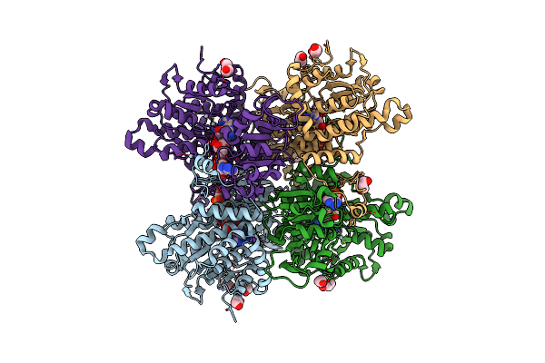
Crystal Structure Of Ciad From Campylobacter Jejuni (C-Terminal Fragment, Orthorhombic P Form)
Organism: Campylobacter jejuni
Method: X-RAY DIFFRACTION Resolution:2.45 Å Release Date: 2023-12-06 Classification: OXIDOREDUCTASE Ligands: BU1, MN, CL |
 |
Organism: Bacillus cereus atcc 14579
Method: X-RAY DIFFRACTION Resolution:2.00 Å Release Date: 2023-07-12 Classification: OXIDOREDUCTASE Ligands: HEM, PGO, BU1, PDO |
 |
Crystal Structure Of Pseudomonas Aeruginosa S-Adenosyl-L-Homocysteine Hydrolase Inhibited By Cd2+ Ions
Organism: Pseudomonas aeruginosa pao1
Method: X-RAY DIFFRACTION Resolution:1.87 Å Release Date: 2023-04-19 Classification: HYDROLASE Ligands: NAD, ADN, BU1, PDO, K, CD, HEZ, CL |
 |
Crystal Structure Of Pseudomonas Aeruginosa S-Adenosyl-L-Homocysteine Hydrolase Inhibited By Hg2+ Ions
Organism: Pseudomonas aeruginosa pao1
Method: X-RAY DIFFRACTION Resolution:1.56 Å Release Date: 2023-04-19 Classification: HYDROLASE Ligands: NAD, ADN, BU1, PDO, K, HG, CL |
 |
Crystal Structure Of Pseudomonas Aeruginosa S-Adenosyl-L-Homocysteine Hydrolase Inhibited By Co2+ Ions.
Organism: Pseudomonas aeruginosa pao1
Method: X-RAY DIFFRACTION Resolution:2.16 Å Release Date: 2023-04-19 Classification: HYDROLASE Ligands: NAD, ADN, PDO, K, CO, BU1, HEZ, CL |
 |
Crystal Structure Of Pseudomonas Aeruginosa S-Adenosyl-L-Homocysteine Hydrolase Inhibited By Zn2+ Ions
Organism: Pseudomonas aeruginosa pao1
Method: X-RAY DIFFRACTION Resolution:1.90 Å Release Date: 2023-04-19 Classification: HYDROLASE Ligands: NAD, ADN, BU1, PG4, K, ZN, BCN, HEZ, PDO, CL |
 |
Plasmodium Falciparum Prolyl-Trna Synthetase (Pfprs) In Complex With L-Proline And Compound L97
Organism: Plasmodium falciparum 3d7
Method: X-RAY DIFFRACTION Resolution:2.39 Å Release Date: 2023-01-04 Classification: LIGASE/LIGASE INHIBITOR Ligands: PRO, 1XK, BU1, CL, HEZ |
 |
Organism: Homo sapiens, Synthetic construct
Method: X-RAY DIFFRACTION Resolution:2.09 Å Release Date: 2022-10-26 Classification: SIGNALING PROTEIN/INHIBITOR Ligands: BU1 |
 |
Plasmodium Falciparum Prolyl-Trna Synthetase (Pfprs) In Complex With Inhibitor L95 And Azetidine
Organism: Plasmodium falciparum
Method: X-RAY DIFFRACTION Resolution:1.94 Å Release Date: 2022-09-07 Classification: LIGASE Ligands: JE6, 02A, EDO, ACT, BU1 |
 |
The Crystal Structure Of The Immature Holo-Enzyme Of Homoserine Dehydrogenase Complexed With Nadp And 1,4-Butandiol From The Hyperthermophilic Archaeon Sulfurisphaera Tokodaii.
Organism: Sulfurisphaera tokodaii (strain dsm 16993 / jcm 10545 / nbrc 100140 / 7)
Method: X-RAY DIFFRACTION Resolution:1.90 Å Release Date: 2022-06-22 Classification: OXIDOREDUCTASE Ligands: NAP, BU1 |
 |
The Crystal Structure Of The K38A/K137A/K233A/K234A Quadruple Mutant Of E. Coli Yggs In Complex With Plp
Organism: Escherichia coli
Method: X-RAY DIFFRACTION Resolution:2.30 Å Release Date: 2022-03-23 Classification: PROTEIN TRANSPORT Ligands: PLP, BU1 |
 |
Crystal Structure Of Probable Gtp-Binding Protein Engb Bound To Gdp From Klebsiella Pneumoniae Subsp. Pneumoniae
Organism: Klebsiella pneumoniae subsp. pneumoniae (strain hs11286)
Method: X-RAY DIFFRACTION Resolution:1.50 Å Release Date: 2021-12-15 Classification: CELL CYCLE Ligands: GDP, GOL, BU1, PGR |
 |
Crystal Structure Of Phosphatidylglycerol Phosphate Synthase In Complex With Phosphatidylglycerol Phosphate
Organism: Staphylococcus aureus (strain n315)
Method: X-RAY DIFFRACTION Resolution:2.50 Å Release Date: 2021-12-08 Classification: TRANSFERASE Ligands: PO9, BU1, OLC, ZN, ACY |
 |
Crystal Structure Of Phosphatidylglycerol Phosphate Synthase In Complex With Cytidine Diphosphate-Diacylglycerol
Organism: Staphylococcus aureus (strain n315)
Method: X-RAY DIFFRACTION Resolution:3.00 Å Release Date: 2021-12-08 Classification: TRANSFERASE Ligands: ZN, 58A, G3P, OLC, BU1, ACY |
 |
Organism: Hypoxylon sp. ec38
Method: X-RAY DIFFRACTION Resolution:1.30 Å Release Date: 2021-09-15 Classification: OXIDOREDUCTASE Ligands: MG, NAG, HEM, IMD, BU1, PEG |
 |
Thermus Thermophilus Transcription Initiation Complex Containing A Template-Strand Purine At Position Tss-2, Gpg Rna Primer, And Cmpcpp
Organism: Thermus thermophilus hb8, Synthetic construct
Method: X-RAY DIFFRACTION Resolution:2.90 Å Release Date: 2021-07-14 Classification: TRANSCRIPTION/DNA/RNA Ligands: MG, BU1, ZN, 2TM |