
Search Count: 14
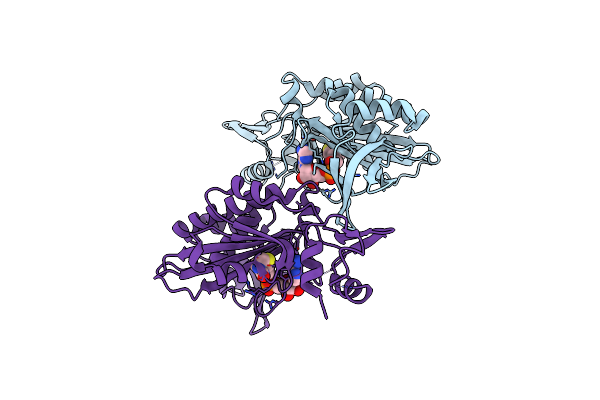 |
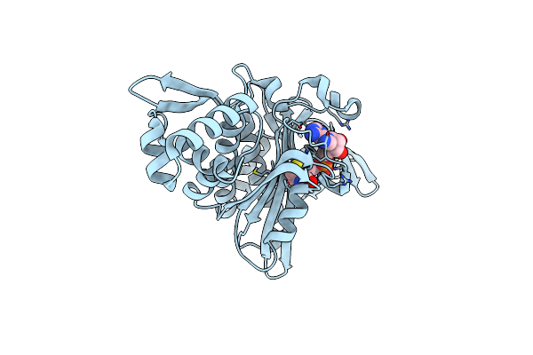
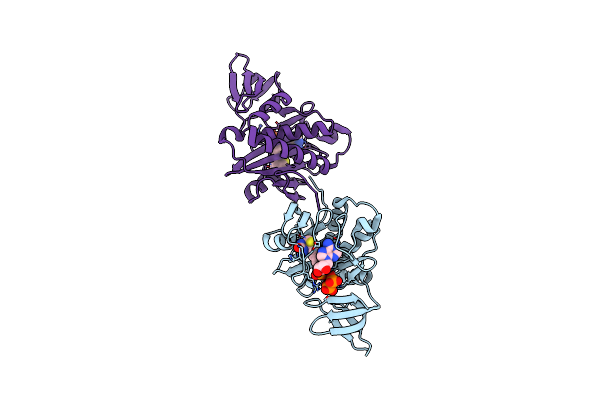
Structural Insights Into Bira From Haemophilus Influenzae, A Bifunctional Protein As A Biotin Protein Ligase And A Transcriptional Repressor
Organism: Haemophilus influenzae rd kw20
Method: X-RAY DIFFRACTION Resolution:2.65 Å Release Date: 2024-09-11 Classification: DNA BINDING PROTEIN Ligands: BT5 |
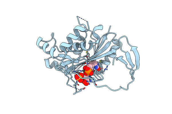 |
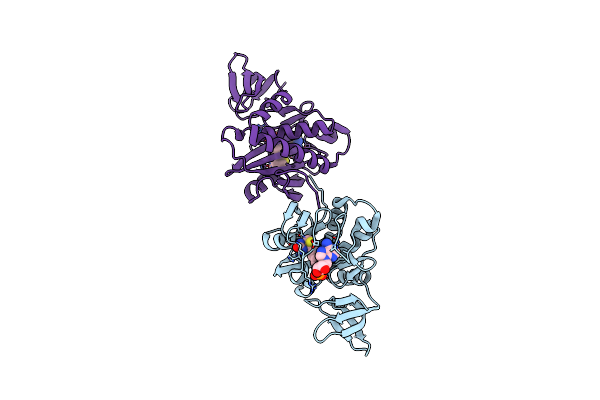
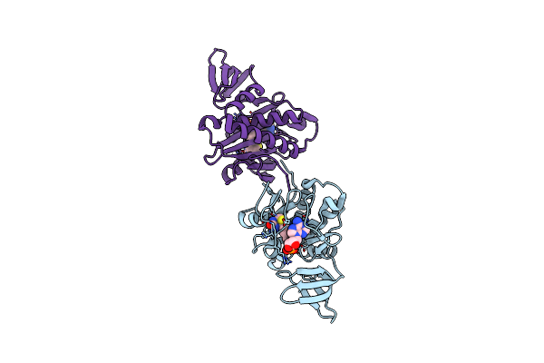
Crystal Structure Of Biotin Protein Ligase From Leishmania Major In Complex With Biotinyl-5-Amp
Organism: Leishmania major
Method: X-RAY DIFFRACTION Resolution:1.97 Å Release Date: 2020-04-08 Classification: LIGASE Ligands: BT5, SO4 |
 |
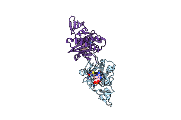
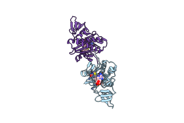
Crystal Structure Of Biotin Protein Ligase (Rv3279C) Of Mycobacterium Tuberculosis, Complexed With Biotinyl-5'-Amp
Organism: Mycobacterium tuberculosis
Method: X-RAY DIFFRACTION Resolution:1.70 Å Release Date: 2014-04-30 Classification: LIGASE Ligands: BT5, SO4 |
 |
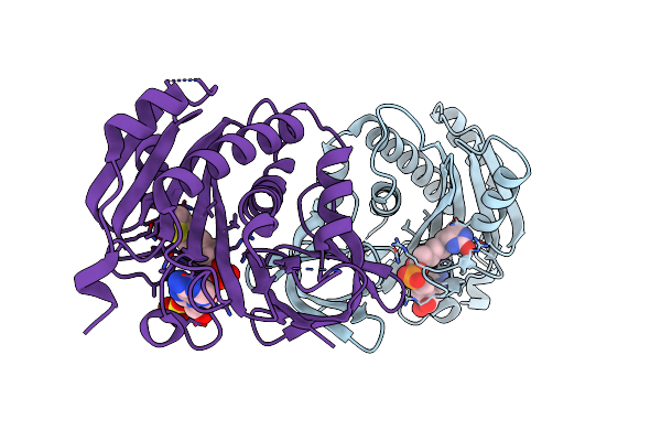
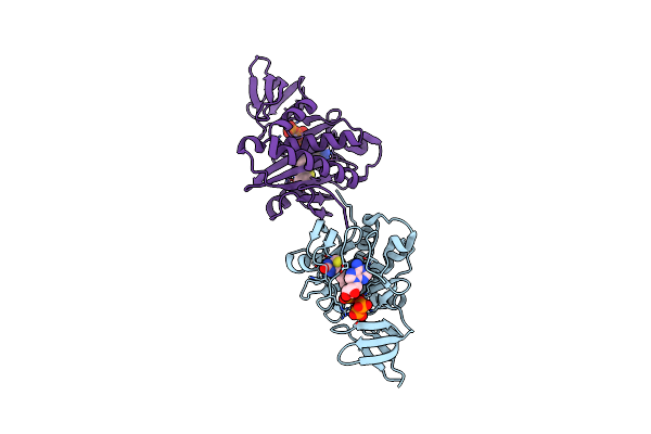
Crystal Structure Of Staphylococcus Aureus Biotin Protein Ligase In Complex With Biotinyl-5'-Amp
Organism: Staphylococcus aureus
Method: X-RAY DIFFRACTION Resolution:2.60 Å Release Date: 2012-12-26 Classification: LIGASE/LIGASE INHIBITOR Ligands: BT5 |
 |
Organism: Staphylococcus aureus
Method: X-RAY DIFFRACTION Resolution:2.60 Å Release Date: 2012-04-18 Classification: LIGASE Ligands: BT5 |
 |
Organism: Staphylococcus aureus
Method: X-RAY DIFFRACTION Resolution:2.50 Å Release Date: 2012-04-18 Classification: LIGASE Ligands: BT5 |
 |
Crystal Structure Of Biotin Protein Ligase From Pyrococcus Horikoshii Complexed With Biotinyl-5'-Amp, Mutation D104A
Organism: Pyrococcus horikoshii
Method: X-RAY DIFFRACTION Resolution:1.84 Å Release Date: 2007-03-26 Classification: LIGASE Ligands: BT5 |
 |
Crystal Structure Of Biotin Protein Ligase From Pyrococcus Horikoshii Complexed With Biotinyl-5'-Amp, Mutation R48A
Organism: Pyrococcus horikoshii
Method: X-RAY DIFFRACTION Resolution:1.28 Å Release Date: 2007-03-01 Classification: LIGASE Ligands: BT5 |
 |
Crystal Structure Of Biotin Protein Ligase From Pyrococcus Horikoshii Ot3 In Complex With Biotinyl-5'-Amp, Mutation Arg51Ala
Organism: Pyrococcus horikoshii
Method: X-RAY DIFFRACTION Resolution:1.60 Å Release Date: 2007-01-31 Classification: LIGASE Ligands: BT5 |
 |
Crystal Structure Of Biotin Protein Ligase From Pyrococcus Horikoshii Ot3 In Complex With Biotinyl-5'-Amp, Pyrophosphate And Mn(2+)
Organism: Pyrococcus horikoshii
Method: X-RAY DIFFRACTION Resolution:2.20 Å Release Date: 2007-01-12 Classification: LIGASE Ligands: MN, POP, BT5 |
 |
Crystal Structure Of Biotin Protein Ligase From Pyrococcus Horikoshii Ot3 In Complex With Biotinyl-5'-Amp, Pyrophosphate And Mg(2+)
Organism: Pyrococcus horikoshii
Method: X-RAY DIFFRACTION Resolution:2.00 Å Release Date: 2006-10-11 Classification: LIGASE Ligands: MG, POP, BT5 |
 |
Crystal Structure Of Biotin Protein Ligase From Pyrococcus Horikoshii Ot3 In Complex With Biotinyl-5'-Amp, K111A Mutation
Organism: Pyrococcus horikoshii
Method: X-RAY DIFFRACTION Resolution:1.85 Å Release Date: 2006-10-06 Classification: LIGASE Ligands: BT5 |
 |
Crystal Structure Of Biotin Protein Ligase From Pyrococcus Horikoshii Ot3 In Complex With Biotinyl-5'-Amp, K111G Mutation
Organism: Pyrococcus horikoshii
Method: X-RAY DIFFRACTION Resolution:1.85 Å Release Date: 2006-08-16 Classification: LIGASE Ligands: BT5 |
 |
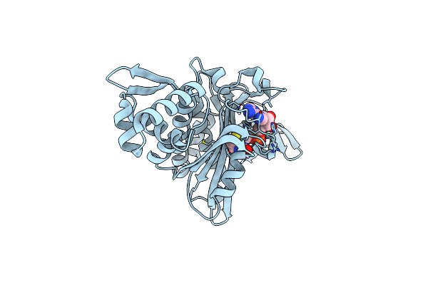
Crystal Structure Of Biotin Protein Ligase From Pyrococcus Horikoshii Ot3 In Complex With Biotinyl-5-Amp
Organism: Pyrococcus horikoshii
Method: X-RAY DIFFRACTION Resolution:1.45 Å Release Date: 2005-10-04 Classification: LIGASE Ligands: BT5 |