
Search Count: 15
 |
Organism: Btrs-betacov/yn2013
Method: ELECTRON MICROSCOPY Release Date: 2025-02-05 Classification: VIRAL PROTEIN Ligands: NAG, BLR |
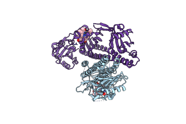 |
Crystal Structure Of Fidcb, A Dual-Cysteine Cyanobacterial Phytochrome Of Fischerella Sp. Pcc 9605
Organism: Fischerella sp. pcc 9605
Method: X-RAY DIFFRACTION Resolution:3.35 Å Release Date: 2025-01-22 Classification: SIGNALING PROTEIN Ligands: BLR |
 |
Organism: Homo sapiens
Method: ELECTRON MICROSCOPY Release Date: 2023-09-13 Classification: MEMBRANE PROTEIN Ligands: BLR, NAG |
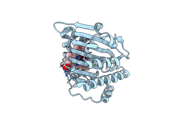 |
Neutron Structure Of Pcya I86D Mutant Complexed With Biliverdin At Room Temperature
Organism: Synechocystis sp. pcc 6803
Method: X-RAY DIFFRACTION, NEUTRON DIFFRACTION Resolution:1.90 Å, 2.0 Å Release Date: 2023-01-25 Classification: OXIDOREDUCTASE Ligands: BLR |
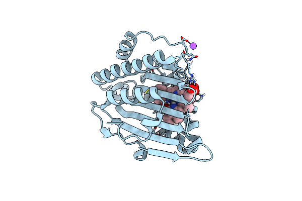 |
Neutron Structure Of Pcya D105N Mutant Complexed With Biliverdin At Room Temperature
Organism: Synechocystis sp. pcc 6803 substr. kazusa
Method: X-RAY DIFFRACTION, NEUTRON DIFFRACTION Resolution:1.38 Å, 2.10 Å Release Date: 2023-01-25 Classification: OXIDOREDUCTASE Ligands: BLR, NA |
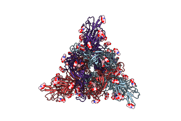 |
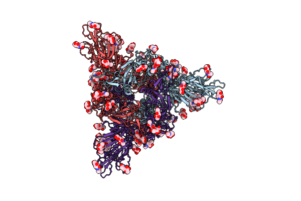
Organism: Severe acute respiratory syndrome coronavirus 2
Method: ELECTRON MICROSCOPY Release Date: 2023-01-04 Classification: VIRAL PROTEIN Ligands: NAG, EIC, BLR |
 |
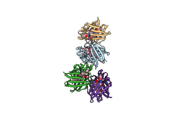
Sars-Cov-2 Spike Glycoprotein Trimer In Closed State After Treatment With Cathepsin L
Organism: Severe acute respiratory syndrome coronavirus 2
Method: ELECTRON MICROSCOPY Release Date: 2023-01-04 Classification: VIRAL PROTEIN Ligands: NAG, BLR |
 |
Structure Of The Chromophore Binding Domain Of Stigmatella Aurantiaca Phytochrome P1, Wild-Type
Organism: Stigmatella aurantiaca dw4/3-1
Method: X-RAY DIFFRACTION Resolution:1.85 Å Release Date: 2018-09-19 Classification: SIGNALING PROTEIN Ligands: BLR |
 |
The Structure Of The Stigmatella Aurantiaca Phytochrome Chromophore Binding Domain T289H Mutant
Organism: Stigmatella aurantiaca dw4/3-1
Method: X-RAY DIFFRACTION Resolution:1.92 Å Release Date: 2018-09-19 Classification: SIGNALING PROTEIN Ligands: BLR |
 |
Organism: Stigmatella aurantiaca dw4/3-1
Method: X-RAY DIFFRACTION Resolution:2.18 Å Release Date: 2018-09-19 Classification: SIGNALING PROTEIN Ligands: BLR |
 |
Organism: Stigmatella aurantiaca dw4/3-1
Method: X-RAY DIFFRACTION Resolution:2.65 Å Release Date: 2018-09-19 Classification: SIGNALING PROTEIN Ligands: BLR |
 |
Stigmatella Aurantiaca Bacterial Phytochrome P1, Pas-Gaf-Phy T289H Mutant, Room Temperature Structure
Organism: Stigmatella aurantiaca dw4/3-1
Method: X-RAY DIFFRACTION Resolution:3.15 Å Release Date: 2018-09-19 Classification: SIGNALING PROTEIN Ligands: BLR |
 |
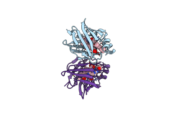
Organism: Anguilla japonica
Method: X-RAY DIFFRACTION Resolution:1.20 Å Release Date: 2013-06-19 Classification: FLUORESCENT PROTEIN Ligands: BLR, PEG |
 |
Organism: Anguilla japonica
Method: X-RAY DIFFRACTION Resolution:2.00 Å Release Date: 2013-06-19 Classification: FLUORESCENT PROTEIN Ligands: BLR |
 |
Organism: Anguilla japonica
Method: X-RAY DIFFRACTION Resolution:2.30 Å Release Date: 2013-06-19 Classification: FLUORESCENT PROTEIN Ligands: BLR |