
Search Count: 106
 |
Organism: Homo sapiens
Method: X-RAY DIFFRACTION Resolution:1.80 Å Release Date: 2026-01-28 Classification: DE NOVO PROTEIN Ligands: BLA |
 |
Organism: Sarbecovirus
Method: ELECTRON MICROSCOPY Release Date: 2025-10-29 Classification: VIRAL PROTEIN Ligands: BLA, NAG, EIC |
 |
Organism: Sarbecovirus
Method: ELECTRON MICROSCOPY Resolution:2.30 Å Release Date: 2025-10-29 Classification: VIRAL PROTEIN Ligands: NAG, BLA |
 |
Organism: Stigmatella aurantiaca
Method: X-RAY DIFFRACTION Resolution:2.30 Å Release Date: 2025-10-08 Classification: SIGNALING PROTEIN Ligands: BLA, BEN |
 |
Organism: Stigmatella aurantiaca
Method: X-RAY DIFFRACTION Resolution:2.30 Å Release Date: 2025-10-08 Classification: SIGNALING PROTEIN Ligands: BLA, BEN |
 |
Organism: Bat sars-like coronavirus khosta-2
Method: ELECTRON MICROSCOPY Release Date: 2025-08-20 Classification: VIRAL PROTEIN Ligands: NAG, BLA, EIC |
 |
Organism: Kenya bat coronavirus btky72
Method: ELECTRON MICROSCOPY Release Date: 2025-02-05 Classification: VIRAL PROTEIN Ligands: NAG, BLA |
 |
Organism: Sarbecovirus
Method: ELECTRON MICROSCOPY Release Date: 2025-02-05 Classification: VIRAL PROTEIN Ligands: NAG, BLA, EIC |
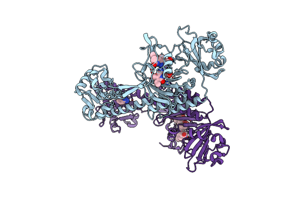 |
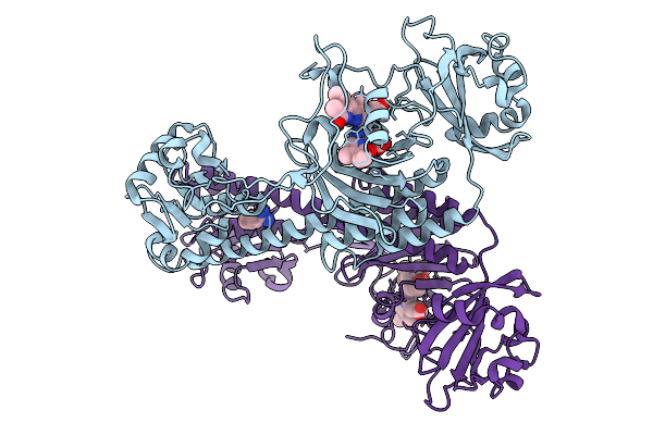
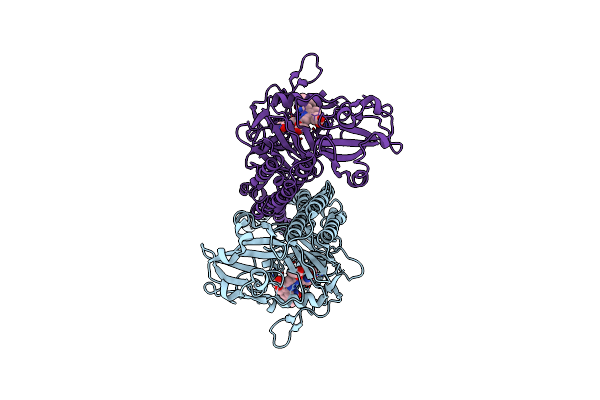
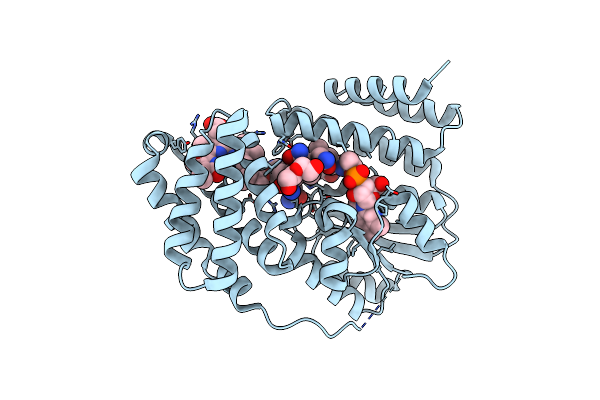
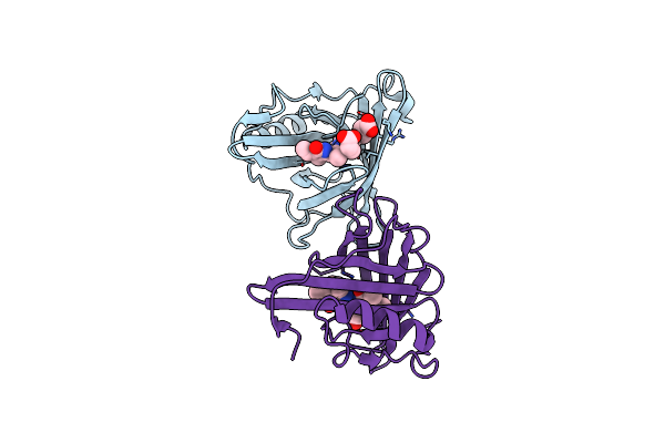
Stigmatella Aurantica Bacteriophytochrome Protein 2 (Sabphp2), Photosensory Core Module, Investigated At Esrf(Ebs) Id29. Dark Structure.
Organism: Stigmatella aurantiaca
Method: X-RAY DIFFRACTION Resolution:2.10 Å Release Date: 2025-01-15 Classification: SIGNALING PROTEIN Ligands: BLA, BEN |
 |
Organism: Stigmatella aurantiaca
Method: ELECTRON MICROSCOPY Release Date: 2024-09-25 Classification: SIGNALING PROTEIN Ligands: BLA |
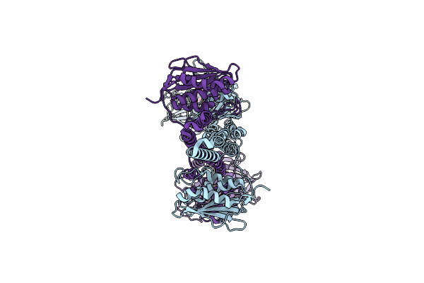 |
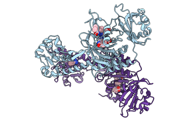
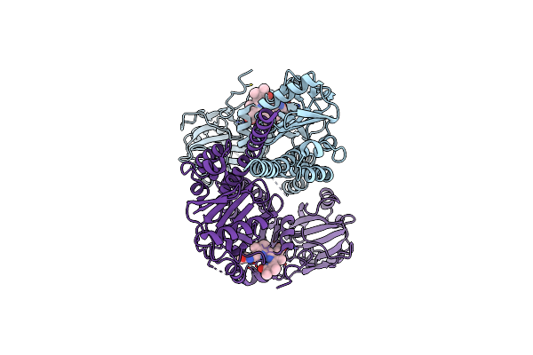
Pr/Pfr Heterodimer (Hybrid) State Of Stigmatella Aurantiaca Bacteriophytochrome 2
Organism: Stigmatella aurantiaca
Method: ELECTRON MICROSCOPY Release Date: 2024-09-25 Classification: SIGNALING PROTEIN Ligands: BLA |
 |
Organism: Stigmatella aurantiaca
Method: ELECTRON MICROSCOPY Release Date: 2024-09-25 Classification: SIGNALING PROTEIN Ligands: BLA |
 |
Organism: Saccharothrix syringae
Method: X-RAY DIFFRACTION Resolution:1.70 Å Release Date: 2024-04-10 Classification: UNKNOWN FUNCTION Ligands: B12, 5AD, BLA, DIO, PEG |
 |
Organism: Saccharothrix syringae
Method: X-RAY DIFFRACTION Resolution:1.98 Å Release Date: 2024-04-10 Classification: UNKNOWN FUNCTION Ligands: B12, BLA, PEG |
 |
Organism: Deinococcus radiodurans r1
Method: ELECTRON MICROSCOPY Release Date: 2022-12-21 Classification: TRANSFERASE Ligands: BLA |
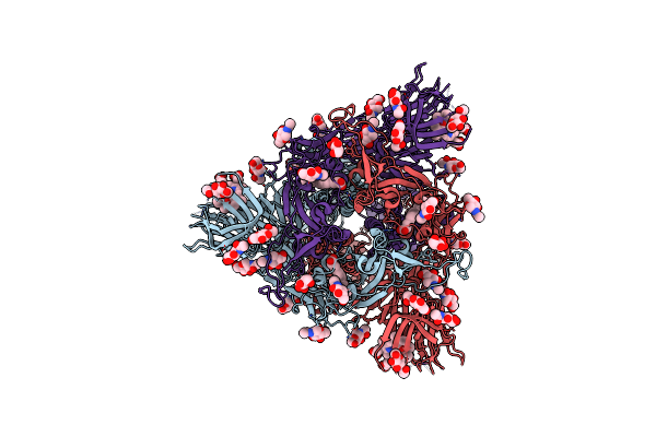 |
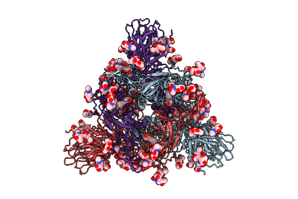
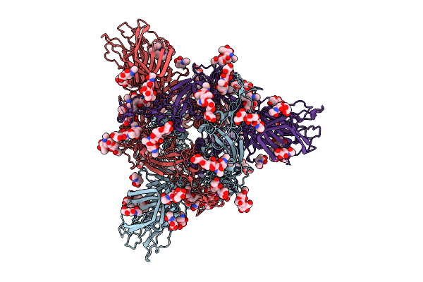
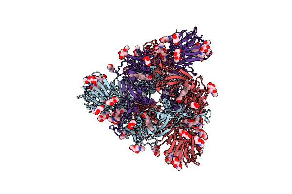
Structure Of Sars-Cov-1 Spike Protein With Engineered X1 Disulfide (S370C And D967C), Locked-1 Conformation
Organism: Severe acute respiratory syndrome coronavirus
Method: ELECTRON MICROSCOPY Release Date: 2022-11-09 Classification: VIRAL PROTEIN Ligands: EIC, NAG, BLA |
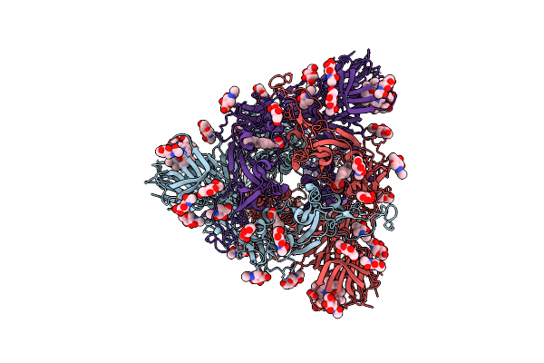 |
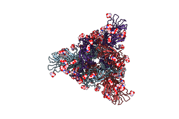
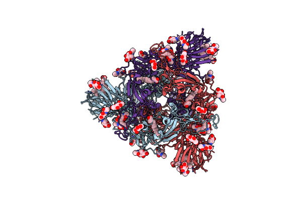
Structure Of Sars-Cov-1 Spike Protein With Engineered X1 Disulfide (S370C And D967C), Locked-112 Conformation
Organism: Severe acute respiratory syndrome coronavirus
Method: ELECTRON MICROSCOPY Release Date: 2022-11-09 Classification: VIRAL PROTEIN Ligands: EIC, NAG, BLA |
 |
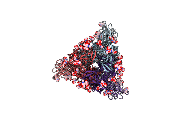
Structure Of Sars-Cov-1 Spike Protein With Engineered X1 Disulfide (S370C And D967C), Locked-122 Conformation
Organism: Severe acute respiratory syndrome coronavirus
Method: ELECTRON MICROSCOPY Release Date: 2022-11-09 Classification: VIRAL PROTEIN Ligands: NAG, BLA, EIC |
 |
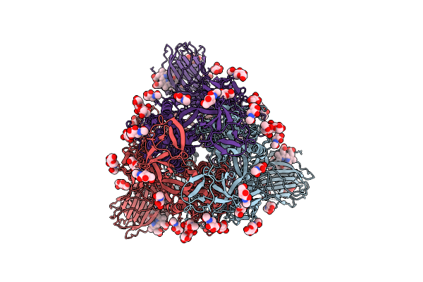
Structure Of Sars-Cov-1 Spike Protein With Engineered X1 Disulfide (S370C And D967C), Locked-2 Conformation
Organism: Severe acute respiratory syndrome coronavirus
Method: ELECTRON MICROSCOPY Release Date: 2022-10-19 Classification: VIRAL PROTEIN Ligands: NAG, BLA, EIC |
 |
Organism: Sander
Method: X-RAY DIFFRACTION Resolution:2.65 Å Release Date: 2022-07-27 Classification: FLUORESCENT PROTEIN Ligands: BLA |