
Search Count: 18
 |
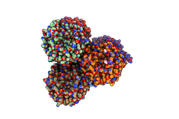
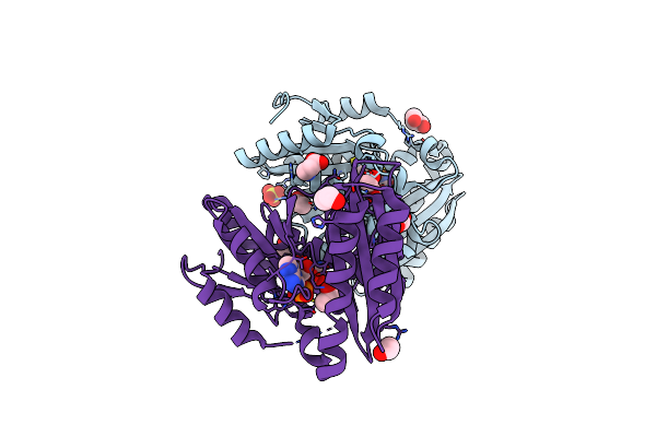
Organism: Homo sapiens
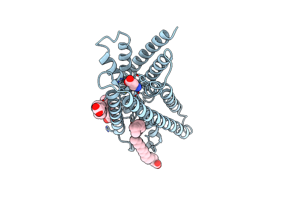
Method: ELECTRON MICROSCOPY Release Date: 2025-12-03 Classification: MEMBRANE PROTEIN Ligands: BAL, CL, NA |
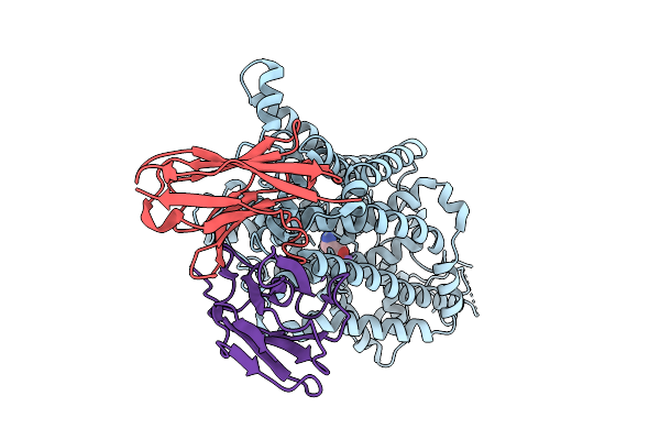 |
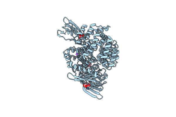
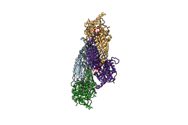
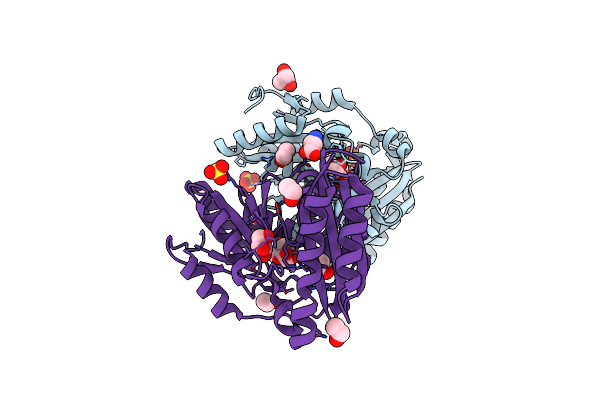
Cryo-Em Structure Of Human Taurine Transporter Taut Bound With Beta-Alanine In An Occluded State
Organism: Homo sapiens
Method: ELECTRON MICROSCOPY Release Date: 2025-08-20 Classification: STRUCTURAL PROTEIN/IMMUNE SYSTEM Ligands: BAL, NA, CL |
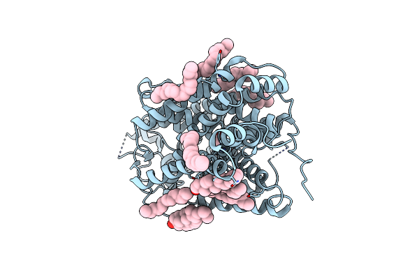 |
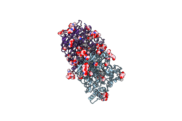
Organism: Homo sapiens
Method: ELECTRON MICROSCOPY Release Date: 2025-04-02 Classification: STRUCTURAL PROTEIN Ligands: BAL, R16, CLR, CL |
 |
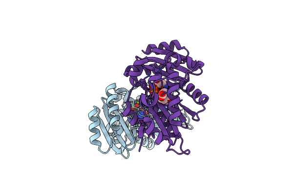
Organism: Homo sapiens
Method: X-RAY DIFFRACTION Resolution:2.25 Å Release Date: 2024-10-23 Classification: TRANSFERASE Ligands: 6CI, BAL |
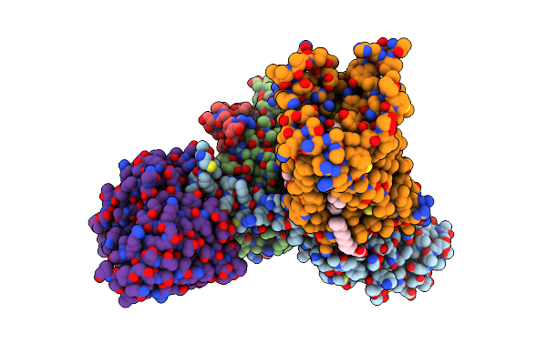 |
Organism: Homo sapiens, Mus musculus, Escherichia coli, Synthetic construct
Method: ELECTRON MICROSCOPY Release Date: 2022-07-20 Classification: SIGNALING PROTEIN Ligands: BAL, PLM |
 |
Organism: Escherichia coli, Homo sapiens
Method: ELECTRON MICROSCOPY Release Date: 2022-07-20 Classification: SIGNALING PROTEIN Ligands: BAL, PLM |
 |
Organism: Acidithiobacillus ferrooxidans
Method: X-RAY DIFFRACTION Resolution:1.95 Å Release Date: 2020-11-11 Classification: HYDROLASE Ligands: SO4, BAL, GLY |
 |
Organism: Escherichia coli k-12
Method: X-RAY DIFFRACTION Resolution:2.80 Å Release Date: 2016-03-02 Classification: HYDROLASE Ligands: ZN, BAL, NA, MLI |
 |
Organism: Homo sapiens
Method: X-RAY DIFFRACTION Resolution:2.50 Å Release Date: 2012-07-04 Classification: HYDROLASE Ligands: BAL, NAG, PG4, EDO, UNX, CL, GLY, PEG |
 |
Crystal Structure Of Pantoate-Beta-Alanine Ligase From Francisella Tularensis Complexed With Beta-Gamma Atp And Beta-Alanine
Organism: Francisella tularensis subsp. tularensis
Method: X-RAY DIFFRACTION Resolution:2.60 Å Release Date: 2011-03-23 Classification: LIGASE Ligands: ANP, PRO, MG, BAL |
 |
Crystal Structure Of Beta-Alanine Synthase From Saccharomyces Kluyveri In Complex With The Product Beta-Alanine
Organism: Saccharomyces kluyveri
Method: X-RAY DIFFRACTION Resolution:2.50 Å Release Date: 2007-10-02 Classification: HYDROLASE Ligands: ZN, BAL, BCN |
 |
Crystal Structure Of A Pantothenate Synthetase Complexed With Amp And Beta-Alanine
Organism: Mycobacterium tuberculosis
Method: X-RAY DIFFRACTION Resolution:1.85 Å Release Date: 2006-02-21 Classification: LIGASE Ligands: SO4, AMP, BAL, GOL, EOH |
 |
Cyclic Peptides Targeting Pdz Domains Of Psd-95: Structural Basis For Enhanced Affinity And Enzymatic Stability
Organism: Rattus norvegicus
Method: SOLUTION NMR Release Date: 2004-05-18 Classification: STRUCTURAL PROTEIN/DE NOVO PROTEIN Ligands: BAL |
 |
Organism: Xenopus laevis, Homo sapiens
Method: X-RAY DIFFRACTION Resolution:2.05 Å Release Date: 2004-05-11 Classification: STRUCTURAL PROTEIN/DNA Ligands: MN, IMT, PYB, ABU, BAL, DIB, OGG, CL |
 |
Crystal Structure Of Pantothenate Synthetase From M. Tuberculosis, Low Occupancy Of Beta-Alanine At The Pantoate Binding Sites
Organism: Mycobacterium tuberculosis
Method: X-RAY DIFFRACTION Resolution:2.10 Å Release Date: 2003-04-23 Classification: LIGASE Ligands: SO4, BAL |
 |
Crystal Structure Of A Pantothenate Synthetase From M. Tuberculosis In Complex With Pantoate
Organism: Mycobacterium tuberculosis
Method: X-RAY DIFFRACTION Resolution:1.80 Å Release Date: 2003-04-22 Classification: LIGASE Ligands: SO4, PAF, BAL, GOL, EOH |
 |
Organism: Xenopus laevis, Synthetic construct
Method: X-RAY DIFFRACTION Resolution:2.30 Å Release Date: 2003-02-18 Classification: STRUCTURAL PROTEIN/DNA Ligands: MN, IMT, PYB, ABU, BAL, DIB |
 |
Organism: Xenopus laevis, Synthetic construct
Method: X-RAY DIFFRACTION Resolution:2.65 Å Release Date: 2003-02-18 Classification: STRUCTURAL PROTEIN/DNA Ligands: MN, IMT, PYB, ABU, BAL, DIB |