
Search Count: 43
 |
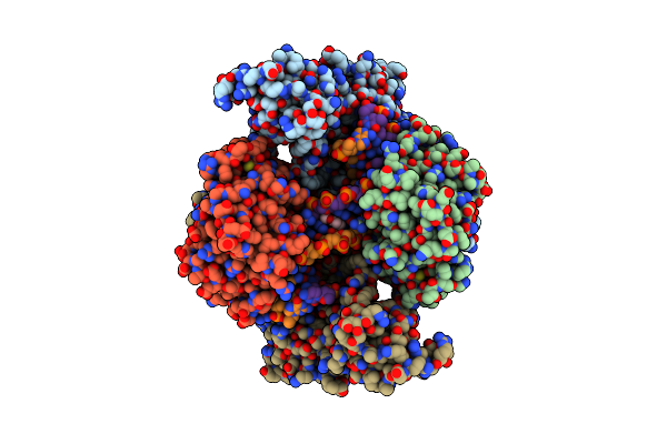
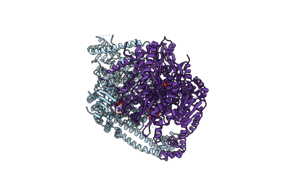
Organism: Mycobacterium tuberculosis, Synthetic construct
Method: ELECTRON MICROSCOPY Release Date: 2025-10-08 Classification: DNA BINDING PROTEIN Ligands: MG, JHN |
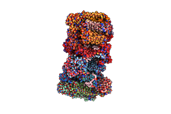 |
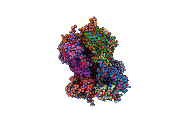
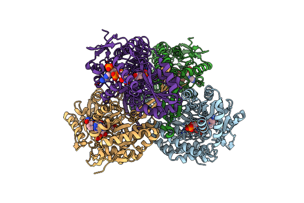
Organism: Mycolicibacterium smegmatis mc2 155
Method: ELECTRON MICROSCOPY Release Date: 2025-04-09 Classification: OXIDOREDUCTASE Ligands: FES, 9YF, PLM, HEM, CDL, A1IRE, HEC, MQ9, 9Y0, CU, HEA, CA, MQ7 |
 |
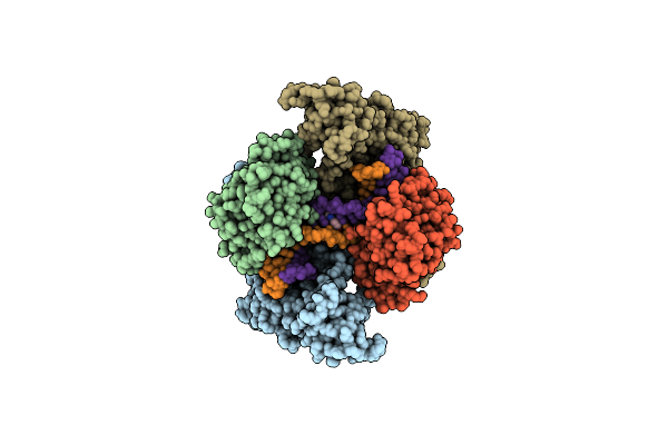
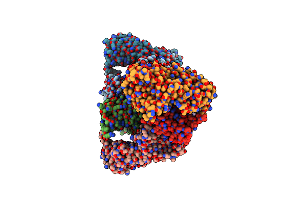
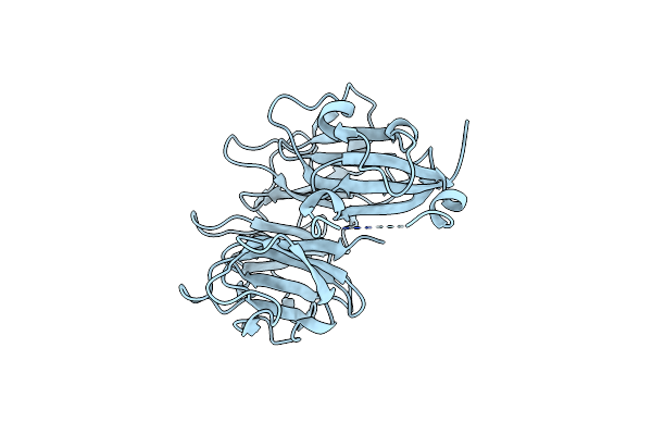
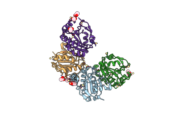
Organism: Mycobacterium tuberculosis, Synthetic construct
Method: ELECTRON MICROSCOPY Release Date: 2025-03-19 Classification: DNA BINDING PROTEIN Ligands: A1H5Q |
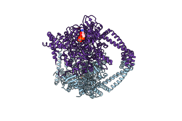 |
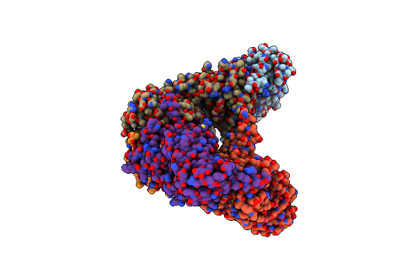
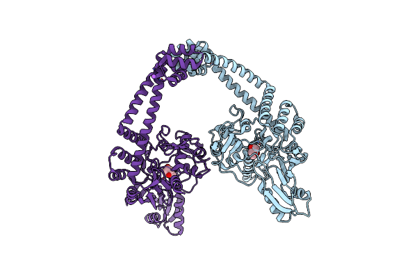
Extremely 'Open' Clamp Structure Of Dna Gyrase: Role Of The Corynebacteriales Gyrb Specific Insert
Organism: Mycobacterium tuberculosis
Method: X-RAY DIFFRACTION Resolution:3.30 Å Release Date: 2019-02-20 Classification: DNA BINDING PROTEIN Ligands: ANP, MG |
 |
Extremely 'Open' Clamp Structure Of Dna Gyrase: Role Of The Corynebacteriales Gyrb Specific Insert
Organism: Mycobacterium tuberculosis
Method: X-RAY DIFFRACTION Resolution:2.60 Å Release Date: 2019-02-20 Classification: DNA BINDING PROTEIN Ligands: MES |
 |
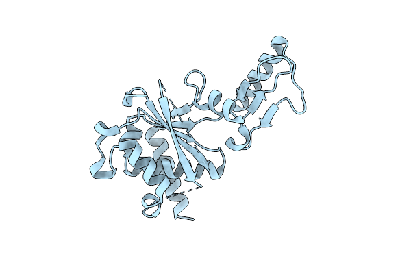
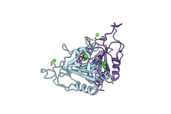
Crystal Structure Of The Atpase Region Of Mycobacterium Tuberculosis Gyrb With Amppnp
Organism: Mycobacterium tuberculosis
Method: X-RAY DIFFRACTION Resolution:2.90 Å Release Date: 2013-09-18 Classification: ISOMERASE Ligands: ANP, MG |
 |
Crystal Structure Of The Atpase Region Of Mycobacterium Tuberculosis Gyrb With Amppnp
Organism: Mycobacterium tuberculosis
Method: X-RAY DIFFRACTION Resolution:2.95 Å Release Date: 2013-09-18 Classification: ISOMERASE Ligands: ANP, MG |
 |
Crystal Structure Of The Atpase Region Of Mycobacterium Tuberculosis Gyrb With Amppcp
Organism: Mycobacterium tuberculosis
Method: X-RAY DIFFRACTION Resolution:3.30 Å Release Date: 2013-09-18 Classification: ISOMERASE Ligands: ACP, MG |
 |
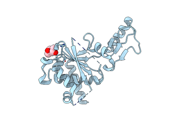
Mycobacterium Tuberculosis Gyrase Type Iia Topoisomerase C-Terminal Domain At 1.4 A Resolution
Organism: Mycobacterium tuberculosis
Method: X-RAY DIFFRACTION Resolution:1.40 Å Release Date: 2013-08-07 Classification: ISOMERASE |
 |
Crystal Structure Of The Second Part Of The Mycobacterium Tuberculosis Dna Gyrase Reaction Core: The Toprim Domain At 1.95 A Resolution
Organism: Mycobacterium tuberculosis
Method: X-RAY DIFFRACTION Resolution:1.95 Å Release Date: 2010-09-29 Classification: ISOMERASE Ligands: MPD |
 |
Crystal Structure Of The First Part Of The Mycobacterium Tuberculosis Dna Gyrase Reaction Core: The Breakage And Reunion Domain At 2.7 A Resolution
Organism: Mycobacterium tuberculosis
Method: X-RAY DIFFRACTION Resolution:2.70 Å Release Date: 2010-07-28 Classification: ISOMERASE Ligands: MPD, NA |
 |
Crystal Structure Of The Second Part Of The Mycobacterium Tuberculosis Dna Gyrase Reaction Core: The Toprim Domain At 2.1 A Resolution
Organism: Mycobacterium tuberculosis
Method: X-RAY DIFFRACTION Resolution:2.10 Å Release Date: 2010-07-28 Classification: ISOMERASE |
 |
Structure Of The Central Domain (Msra) Of Neisseria Meningitidis Pilb (Reduced Form)
Organism: Neisseria meningitidis
Method: X-RAY DIFFRACTION Resolution:2.00 Å Release Date: 2008-02-19 Classification: OXIDOREDUCTASE |
 |
Structure Of The Central Domain (Msra) Of Neisseria Meningitidis Pilb (Complex With A Substrate)
Organism: Neisseria meningitidis
Method: X-RAY DIFFRACTION Resolution:2.24 Å Release Date: 2008-02-19 Classification: OXIDOREDUCTASE Ligands: SSM |
 |
Structure Of The Central Domain (Msra) Of Neisseria Meningitidis Pilb (Sulfenic Acid Form)
Organism: Neisseria meningitidis
Method: X-RAY DIFFRACTION Resolution:2.00 Å Release Date: 2008-02-19 Classification: OXIDOREDUCTASE |
 |
Structure Of The Central Domain (Msra) Of Neisseria Meningitidis Pilb (Oxidized Form)
Organism: Neisseria meningitidis
Method: X-RAY DIFFRACTION Resolution:1.95 Å Release Date: 2008-02-19 Classification: OXIDOREDUCTASE Ligands: ACT |
 |
Thioacylenzyme Intermediate Of Gapn From S. Mutans, New Data Integration And Refinement.
Organism: Streptococcus mutans
Method: X-RAY DIFFRACTION Resolution:2.19 Å Release Date: 2007-12-25 Classification: OXIDOREDUCTASE Ligands: G3H, NAP |
 |
Crystal Structure Of The Poplar Glutathione Peroxidase 5 In The Reduced Form
Organism: Populus trichocarpa x populus deltoides
Method: X-RAY DIFFRACTION Resolution:2.00 Å Release Date: 2007-07-24 Classification: OXIDOREDUCTASE Ligands: CD, ACT |
 |
Crystal Structure Of The Poplar Glutathione Peroxidase 5 In The Oxidized Form
Organism: Populus trichocarpa x populus deltoides
Method: X-RAY DIFFRACTION Resolution:2.45 Å Release Date: 2007-07-24 Classification: OXIDOREDUCTASE Ligands: CA |
 |
Crystal Structure Of Thioacylenzyme Intermediate Of An Nadp Dependent Aldehyde Dehydrogenase
Organism: Streptococcus mutans
Method: X-RAY DIFFRACTION Resolution:2.55 Å Release Date: 2006-05-02 Classification: OXIDOREDUCTASE Ligands: NAP, G3H |