Planned Maintenance: Some services may turn out to be unavailable from 15th January, 2026 to 16th January, 2026. We apologize for the inconvenience!

Planned Maintenance: Some services may turn out to be unavailable from 15th January, 2026 to 16th January, 2026. We apologize for the inconvenience!
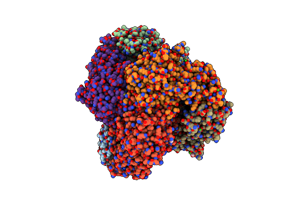 |
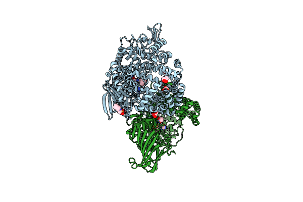
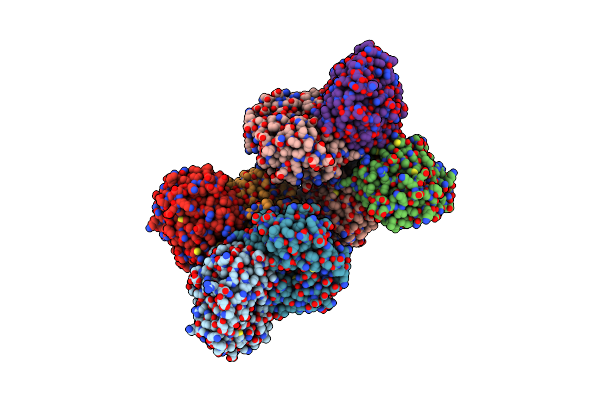
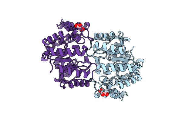
Metal Dependent Activation Of Plasmodium Falciparum M17 Aminopeptidase (Inactive Form), Spacegroup P22121
Organism: Plasmodium falciparum
Method: X-RAY DIFFRACTION Resolution:2.03 Å Release Date: 2022-06-29 Classification: PEPTIDE BINDING PROTEIN Ligands: ZN, CO3, ACT, CA, EDO, PO4, GOL |
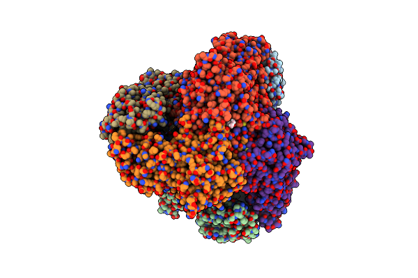 |
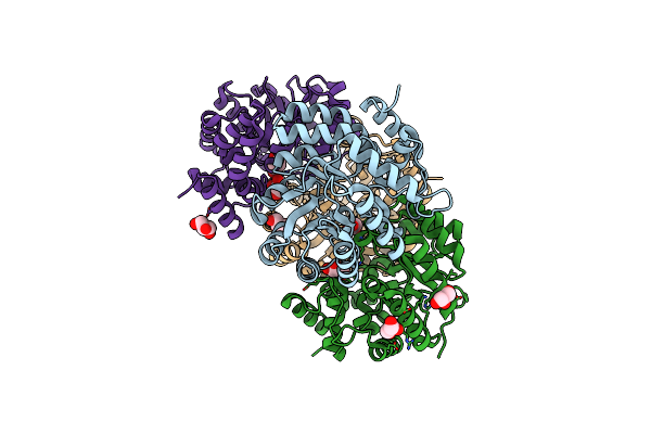
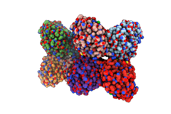
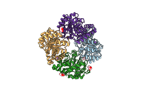
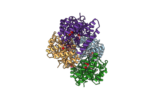
Metal Dependent Activation Of Plasmodium Falciparum M17 Aminopeptidase, Spacegroup P22121 After Crystals Soaked With Zn2+
Organism: Plasmodium falciparum
Method: X-RAY DIFFRACTION Resolution:2.30 Å Release Date: 2022-06-29 Classification: HYDROLASE Ligands: CO3, ZN, MPD, SO4 |
 |
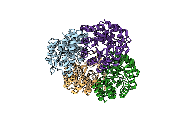
Organism: Plasmodium vivax
Method: X-RAY DIFFRACTION Resolution:2.33 Å Release Date: 2020-12-16 Classification: HYDROLASE Ligands: GOL, PEG, ZN, SO4 |
 |
Organism: Plasmodium vivax
Method: ELECTRON MICROSCOPY Release Date: 2020-12-16 Classification: HYDROLASE Ligands: CO3, MN |
 |
Organism: Anopheles gambiae, Drosophila
Method: X-RAY DIFFRACTION Resolution:2.65 Å Release Date: 2015-06-17 Classification: HYDROLASE Ligands: ZN, GOL, NHE, CU |
 |
Organism: Escherichia coli
Method: X-RAY DIFFRACTION Resolution:2.28 Å Release Date: 2015-03-25 Classification: TRANSFERASE Ligands: SO4 |
 |
Organism: Agrobacterium sp.
Method: X-RAY DIFFRACTION Resolution:1.55 Å Release Date: 2013-12-04 Classification: BIOSYNTHETIC PROTEIN Ligands: GOL |
 |
Organism: Agrobacterium sp.
Method: X-RAY DIFFRACTION Resolution:1.45 Å Release Date: 2013-12-04 Classification: BIOSYNTHETIC PROTEIN |
 |
Organism: Agrobacterium sp.
Method: X-RAY DIFFRACTION Resolution:1.30 Å Release Date: 2013-12-04 Classification: BIOSYNTHETIC PROTEIN Ligands: LYS, GOL |
 |
Dihydrodipicolinate Synthase From The Common Grapevine With Pyruvate And Lysine
Organism: Vitis vinifera
Method: X-RAY DIFFRACTION Resolution:2.40 Å Release Date: 2013-09-04 Classification: HYDROLASE Ligands: LYS |
 |
Organism: Vitis vinifera
Method: X-RAY DIFFRACTION Resolution:2.20 Å Release Date: 2012-07-25 Classification: LYASE Ligands: BR, CL |
 |
Organism: Arabidopsis thaliana
Method: X-RAY DIFFRACTION Resolution:2.00 Å Release Date: 2012-07-18 Classification: LYASE |
 |
The Structure Of Dihydrodipicolinate Synthase 2 From Arabidopsis Thaliana In Complex With (S)-Lysine
Organism: Arabidopsis thaliana
Method: X-RAY DIFFRACTION Resolution:2.20 Å Release Date: 2012-07-18 Classification: LYASE |
 |
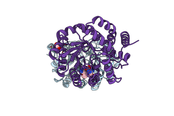
X-Ray Crystal Structure And Specificity Of The Plasmodium Falciparum Malaria Aminopeptidase
Organism: Plasmodium falciparum 3d7
Method: X-RAY DIFFRACTION Resolution:2.60 Å Release Date: 2012-06-27 Classification: HYDROLASE Ligands: ZN |
 |
High-Resolution Structure Of Dhdps From Clostridium Botulinum In Complex With Pyruvate
Organism: Clostridium botulinum a
Method: X-RAY DIFFRACTION Resolution:1.19 Å Release Date: 2010-06-30 Classification: LYASE Ligands: GOL |
 |
Crystal Structure Of Dihydrodipicolinate Synthase From Bacillus Anthracis In Complex With Its Substrate, Pyruvate
Organism: Bacillus anthracis
Method: X-RAY DIFFRACTION Resolution:2.15 Å Release Date: 2009-11-24 Classification: LYASE Ligands: GOL, NA |
 |
Crystal Structure Of Dihydrodipicolinate Synthase From Methicillin-Resistant Staphylococcus Aureus
Organism: Staphylococcus aureus
Method: X-RAY DIFFRACTION Resolution:1.45 Å Release Date: 2008-08-05 Classification: LYASE Ligands: CL, GOL |