Search Count: 18
 |
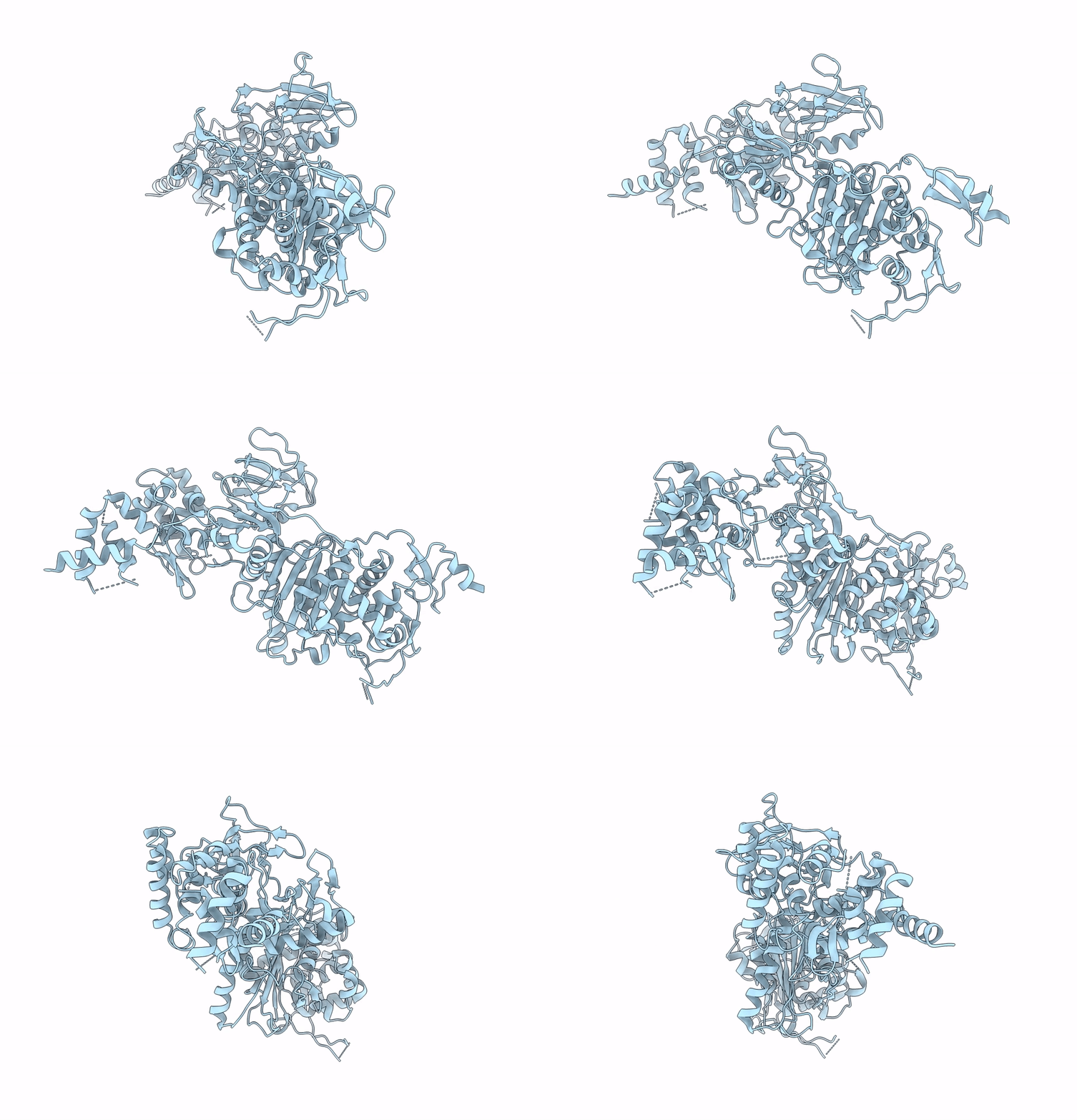
Organism: Homo sapiens
Method: ELECTRON MICROSCOPY Release Date: 2023-02-22 Classification: TRANSFERASE/IMMUNE SYSTEM |
 |
Organism: Escherichia coli (strain k12)
Method: ELECTRON MICROSCOPY Release Date: 2021-05-26 Classification: TRANSFERASE, HYDROLASE |
 |
Organism: Escherichia coli o127:h6 (strain e2348/69 / epec)
Method: ELECTRON MICROSCOPY Release Date: 2020-12-30 Classification: PROTEIN TRANSPORT |
 |
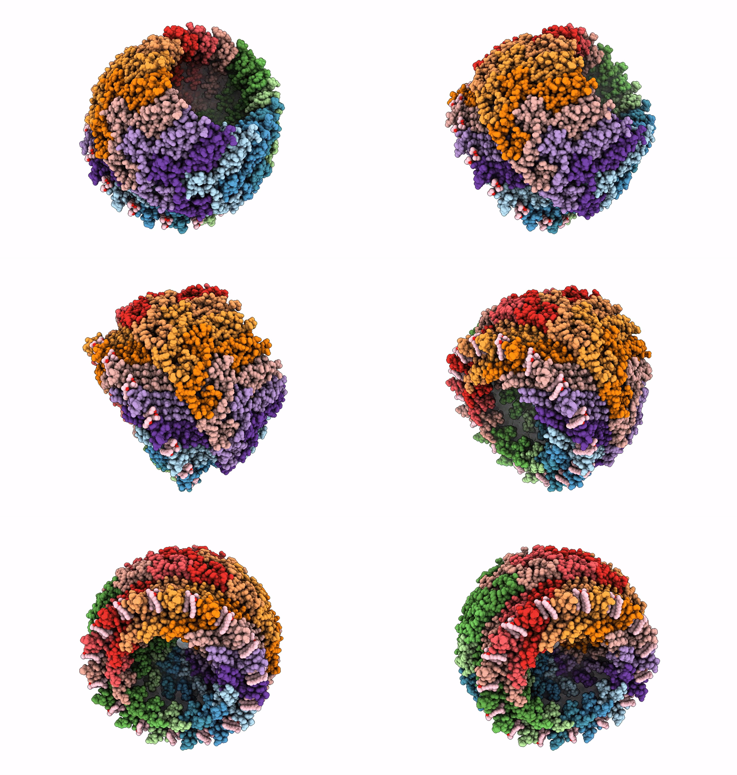
Cryo-Em Structure Of The Nonameric Escv Cytosolic Domain From The Type Iii Secretion System
Organism: Escherichia coli o127:h6 (strain e2348/69 / epec)
Method: ELECTRON MICROSCOPY Release Date: 2020-11-25 Classification: PROTEIN TRANSPORT |
 |
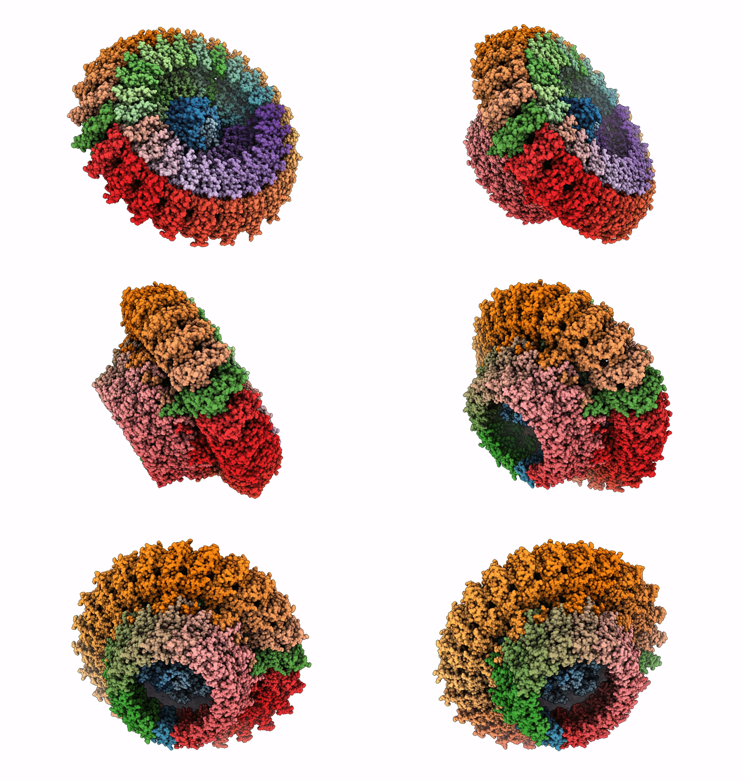
Organism: Salmonella typhimurium (strain lt2 / sgsc1412 / atcc 700720)
Method: ELECTRON MICROSCOPY Release Date: 2019-10-23 Classification: PROTEIN TRANSPORT Ligands: LDA |
 |
Focussed Refinement Of Invgn0N1:Spapqr:Prghk From Salmonella Spi-1 Injectisome Nc-Base
Organism: Salmonella typhimurium (strain lt2 / sgsc1412 / atcc 700720)
Method: ELECTRON MICROSCOPY Release Date: 2019-10-23 Classification: PROTEIN TRANSPORT |
 |
Focussed Refinement Of Invgn0N1:Spapqr:Prgij From The Salmonella Spi-1 Injectisome Needle Complex
Organism: Salmonella typhimurium (strain lt2 / sgsc1412 / atcc 700720)
Method: ELECTRON MICROSCOPY Release Date: 2019-10-23 Classification: PROTEIN TRANSPORT |
 |
Organism: Salmonella typhimurium (strain lt2 / sgsc1412 / atcc 700720)
Method: ELECTRON MICROSCOPY Release Date: 2019-10-23 Classification: PROTEIN TRANSPORT |
 |
Organism: Salmonella typhimurium (strain lt2 / sgsc1412 / atcc 700720)
Method: ELECTRON MICROSCOPY Release Date: 2019-10-23 Classification: PROTEIN TRANSPORT |
 |
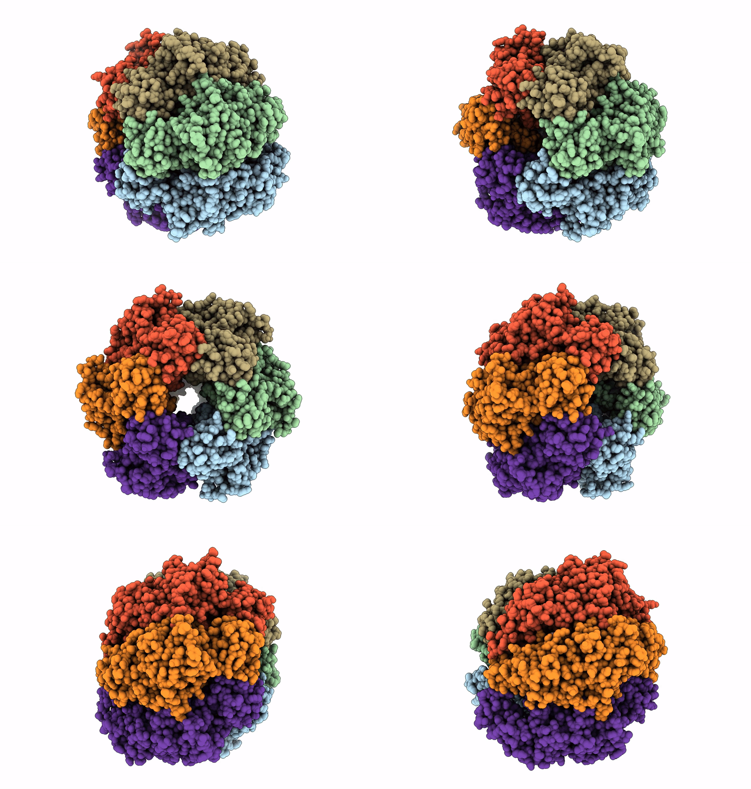
Focussed Refinement Of Invgn0N1:Prghk:Spapqr:Prgij From Salmonella Spi-1 Injectisome Nc-Base
Organism: Salmonella typhimurium (strain lt2 / sgsc1412 / atcc 700720)
Method: ELECTRON MICROSCOPY Release Date: 2019-10-23 Classification: PROTEIN TRANSPORT |
 |
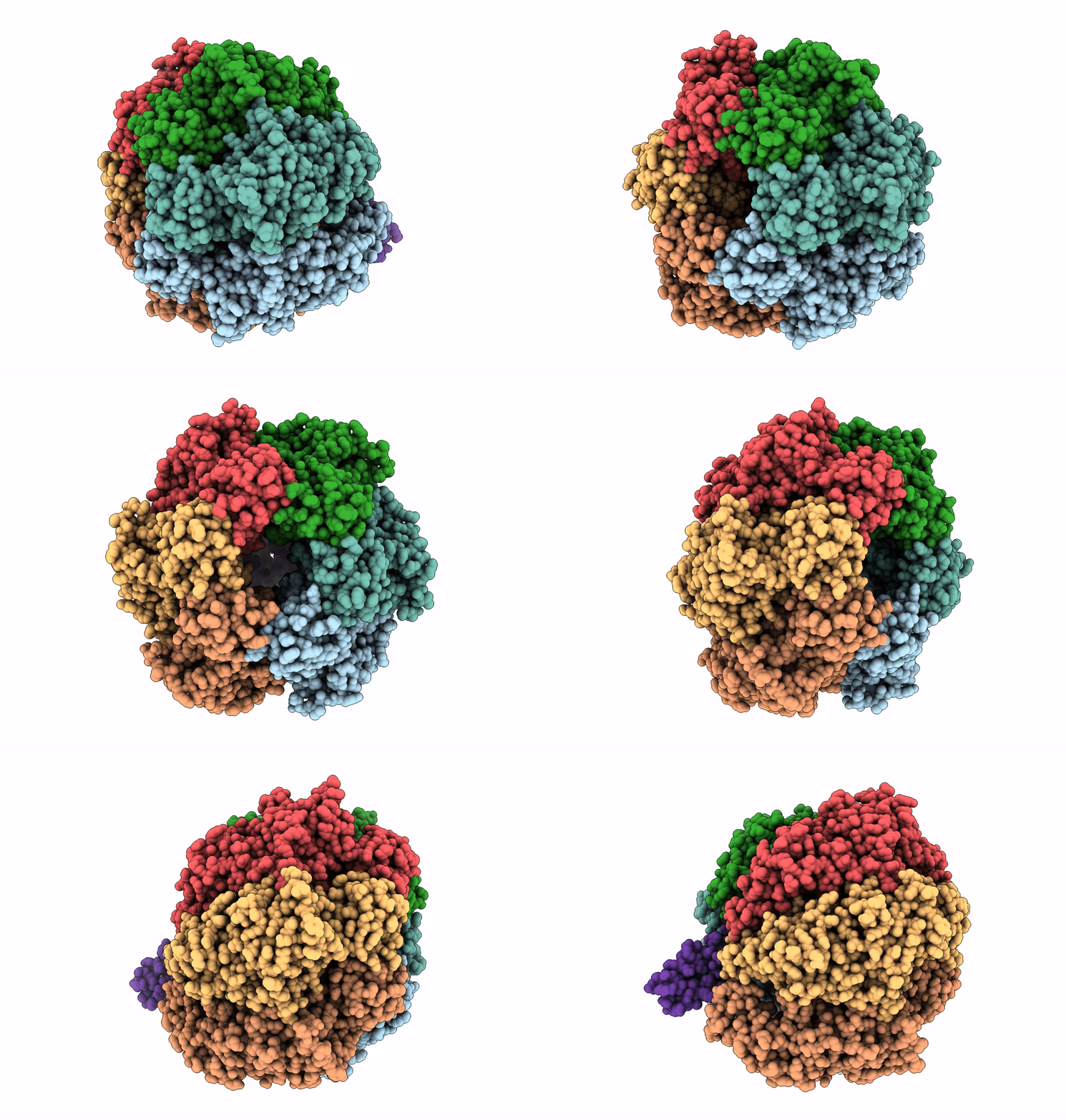
Structure Of The Assembled Atpase Escn From The Enteropathogenic E. Coli (Epec) Type Iii Secretion System
Organism: Escherichia coli o127:h6 (strain e2348/69 / epec)
Method: ELECTRON MICROSCOPY Release Date: 2019-02-20 Classification: HYDROLASE Ligands: ADP, MG, AF3 |
 |
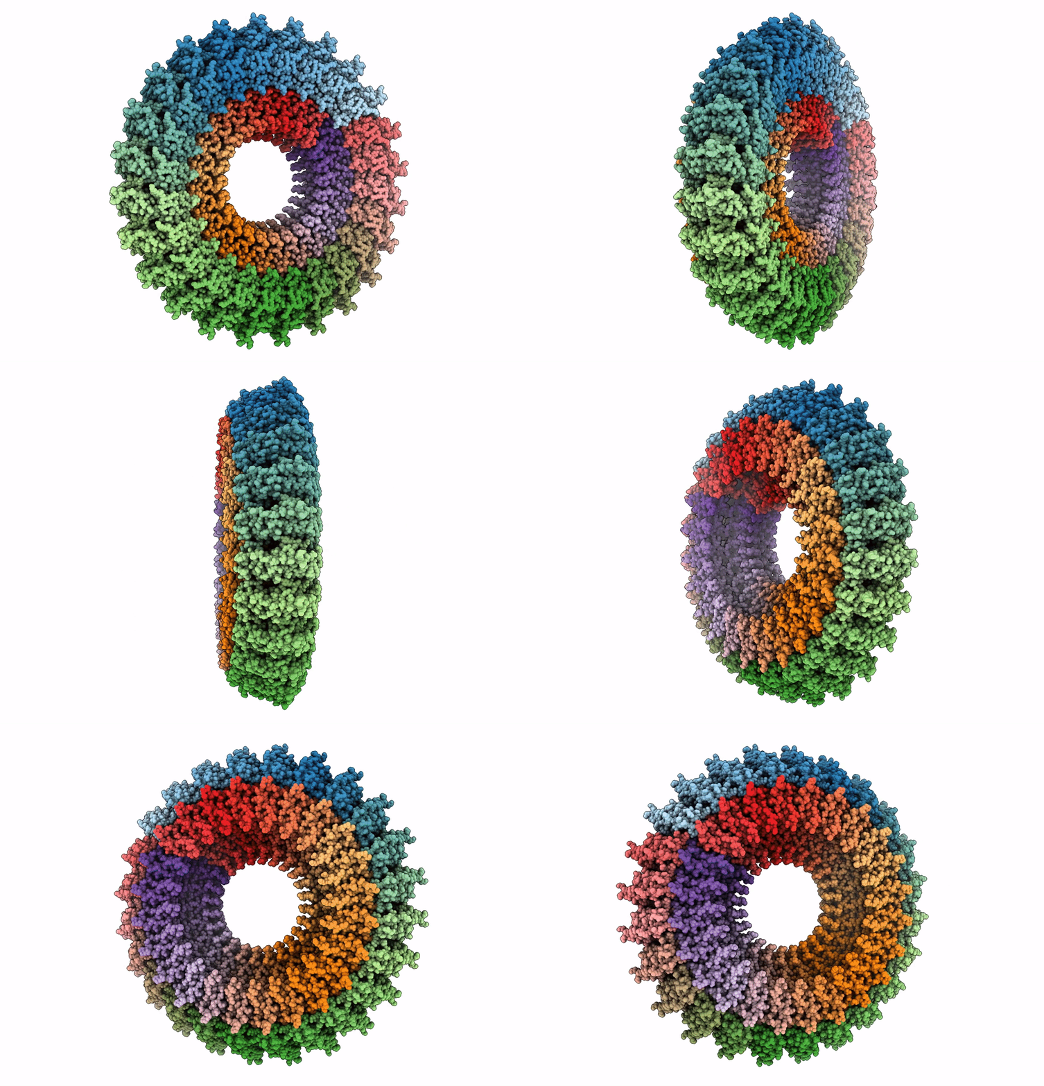
Structure Of The Assembled Atpase Escn In Complex With Its Central Stalk Esco From The Enteropathogenic E. Coli (Epec) Type Iii Secretion System
Organism: Escherichia coli o127:h6 (strain e2348/69 / epec)
Method: ELECTRON MICROSCOPY Release Date: 2019-02-20 Classification: HYDROLASE Ligands: ADP, MG, AF3 |
 |
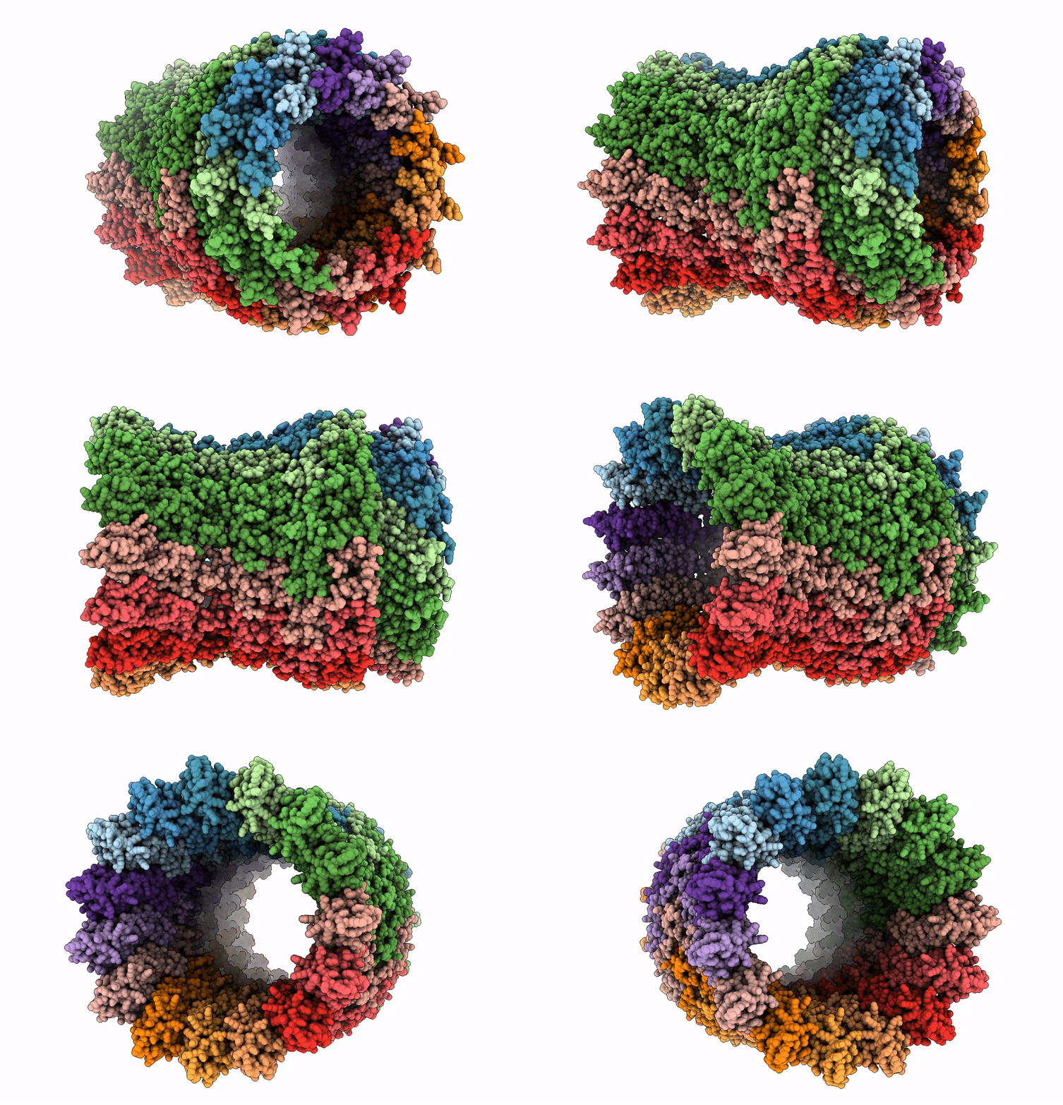
Structure Of The Periplasmic Domains Of Prgh And Prgk From The Assembled Salmonella Type Iii Secretion Injectisome Needle Complex
Organism: Salmonella enterica subsp. enterica serovar typhimurium
Method: ELECTRON MICROSCOPY Release Date: 2018-10-03 Classification: MEMBRANE PROTEIN |
 |
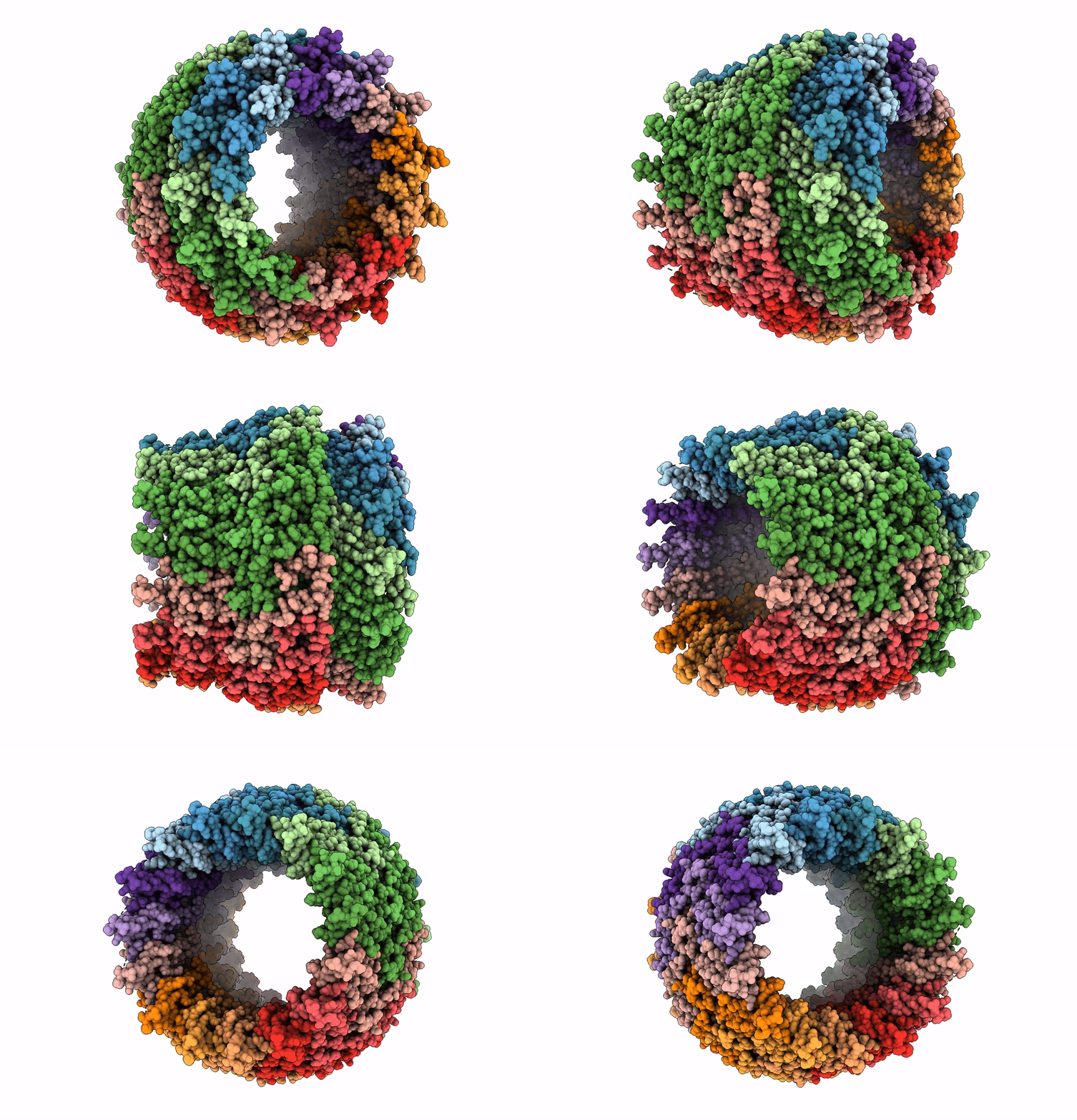
Structure Of The Salmonella Spi-1 Type Iii Secretion Injectisome Secretin Invg In The Open Gate State
Organism: Salmonella enterica subsp. enterica serovar typhimurium
Method: ELECTRON MICROSCOPY Release Date: 2018-10-03 Classification: MEMBRANE PROTEIN |
 |
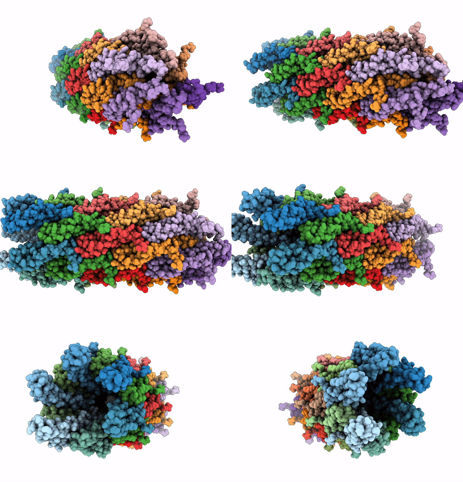
Structure Of The Salmonella Spi-1 Type Iii Secretion Injectisome Secretin Invg (Residues 176-End) In The Open Gate State
Organism: Salmonella enterica subsp. enterica serovar typhimurium
Method: ELECTRON MICROSCOPY Release Date: 2018-10-03 Classification: MEMBRANE PROTEIN |
 |
Structure Of The Salmonella Spi-1 Type Iii Secretion Injectisome Needle Filament
Organism: Salmonella enterica subsp. enterica serovar typhimurium
Method: ELECTRON MICROSCOPY Release Date: 2018-10-03 Classification: MEMBRANE PROTEIN |
 |
Organism: Pseudomonas aeruginosa
Method: X-RAY DIFFRACTION Resolution:1.40 Å Release Date: 2008-09-09 Classification: TRANSPORT PROTEIN |
 |
Organism: Pseudomonas aeruginosa
Method: X-RAY DIFFRACTION Resolution:1.90 Å Release Date: 2008-09-09 Classification: TRANSPORT PROTEIN Ligands: MG |

