
Search Count: 12
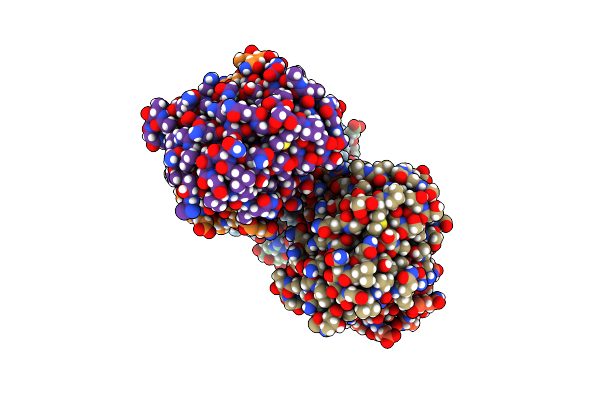 |
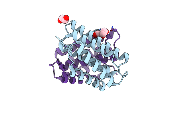
Crystal Structure Of An Asymmetric Dimer Of The N-Terminal Domain Of Euprosthenops Australis Major Ampullate Spidroin 1 (Dragline Silk)
Organism: Euprosthenops australis
Method: X-RAY DIFFRACTION Resolution:2.10 Å Release Date: 2019-07-17 Classification: PROTEIN FIBRIL Ligands: SO4 |
 |
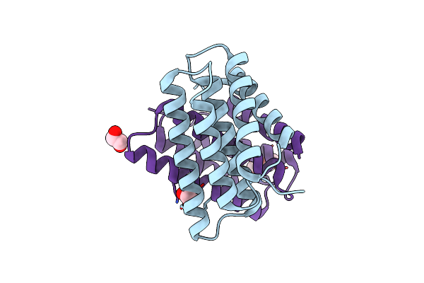
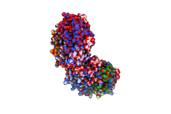
Crystal Structure Of The Thermostable Cellobiohydrolase Cel7A From The Fungus Humicola Grisea Var. Thermoidea.
Organism: Humicola grisea var. thermoidea
Method: X-RAY DIFFRACTION Resolution:1.80 Å Release Date: 2014-09-10 Classification: HYDROLASE Ligands: NAG, PEG |
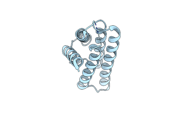 |
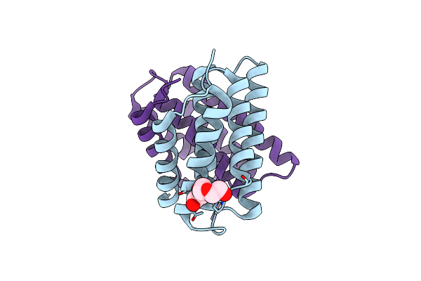
Structure Of Monomeric Nt From Euprosthenops Australis Major Ampullate Spidroin 1 (Masp1)
Organism: Euprosthenops australis
Method: X-RAY DIFFRACTION Resolution:1.70 Å Release Date: 2012-08-01 Classification: STRUCTURAL PROTEIN Ligands: BR |
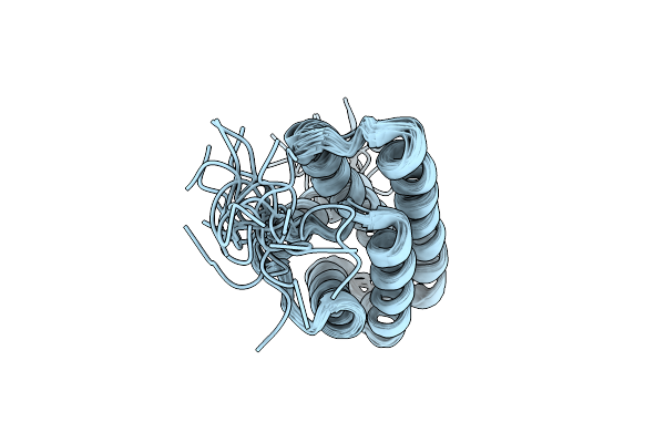 |
Nmr Structure Of A Monomeric Mutant (A72R) Of Major Ampullate Spidroin 1 N-Terminal Domain
Organism: Euprosthenops australis
Method: SOLUTION NMR Release Date: 2012-06-27 Classification: STRUCTURAL PROTEIN |
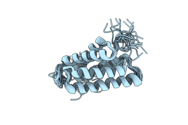 |
Organism: Euprosthenops australis
Method: SOLUTION NMR Release Date: 2012-06-27 Classification: STRUCTURAL PROTEIN |
 |
Organism: Homo sapiens
Method: X-RAY DIFFRACTION Resolution:2.20 Å Release Date: 2012-02-15 Classification: SURFACTANT PROTEIN |
 |
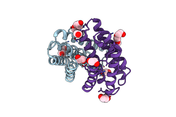
Self-Assembly Of Spider Silk Proteins Is Controlled By A Ph-Sensitive Relay
Organism: Euprosthenops australis
Method: X-RAY DIFFRACTION Resolution:1.70 Å Release Date: 2010-05-12 Classification: STRUCTURAL PROTEIN Ligands: EDO, PEG |
 |
Self-Assembly Of Spider Silk Proteins Is Controlled By A Ph-Sensitive Relay
Organism: Euprosthenops australis
Method: X-RAY DIFFRACTION Resolution:2.20 Å Release Date: 2010-05-12 Classification: STRUCTURAL PROTEIN Ligands: PGE |
 |
Self-Assembly Of Spider Silk Proteins Is Controlled By A Ph-Sensitive Relay
Organism: Euprosthenops australis
Method: X-RAY DIFFRACTION Resolution:2.30 Å Release Date: 2010-05-12 Classification: STRUCTURAL PROTEIN Ligands: EDO, PEG, PGE |
 |
Self-Assembly Of Spider Silk Proteins Is Controlled By A Ph-Sensitive Relay
Organism: Euprosthenops australis
Method: X-RAY DIFFRACTION Resolution:2.15 Å Release Date: 2010-05-12 Classification: STRUCTURAL PROTEIN Ligands: EDO, PGE |
 |
Crystal Structure Of Human Heat-Labile Enterotoxin In Complex With A Blood Group A Antigen Analog
Organism: Escherichia coli
Method: X-RAY DIFFRACTION Resolution:2.53 Å Release Date: 2007-08-14 Classification: TOXIN |
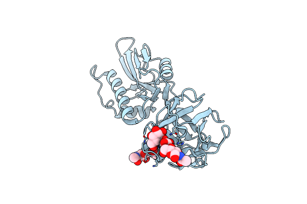 |
Crystal Structure Of Moa, A Lectin From The Mushroom Marasmius Oreades In Complex With The Trisaccharide Gal(1,3)Gal(1,4)Glcnac
Organism: Marasmius oreades
Method: X-RAY DIFFRACTION Resolution:2.41 Å Release Date: 2007-05-22 Classification: SUGAR BINDING PROTEIN |