Planned Maintenance: Some services may turn out to be unavailable from 15th January, 2026 to 16th January, 2026. We apologize for the inconvenience!

Planned Maintenance: Some services may turn out to be unavailable from 15th January, 2026 to 16th January, 2026. We apologize for the inconvenience!
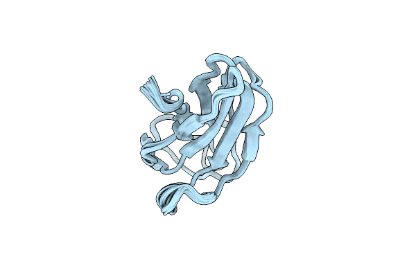 |
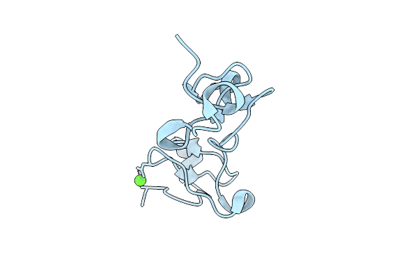
Solution Structure Of The Silkworm Bgrp/Gnbp3 N-Terminal Domain Reveals The Mechanism For B-1,3-Glucan Specific Recognition
Organism: Bombyx mori
Method: SOLUTION NMR Release Date: 2009-06-23 Classification: SUGAR BINDING PROTEIN |
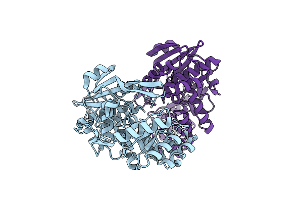 |
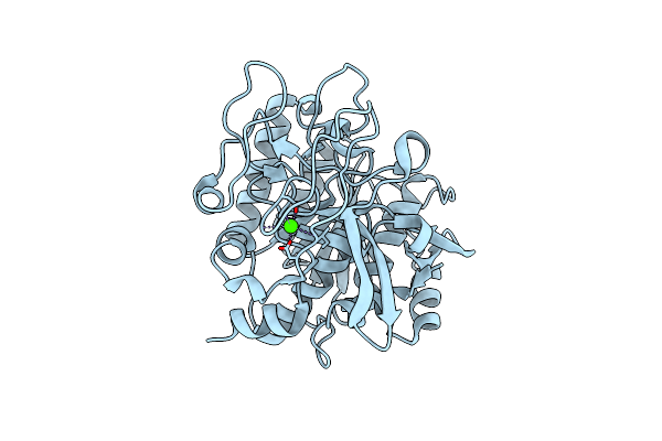
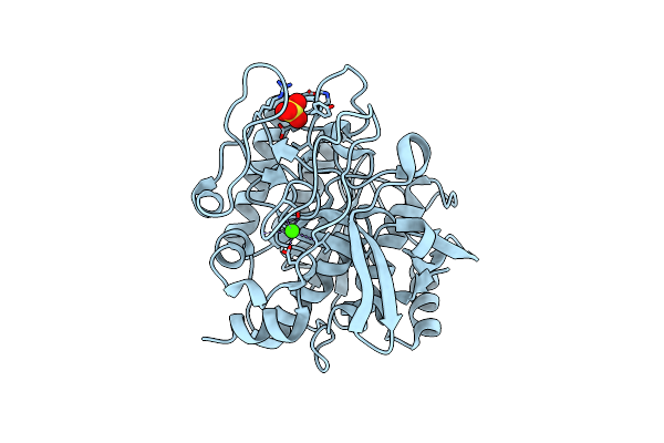
Crystal Structure Of N-Acylamino Acid Racemase From Thermus Thermophilus Hb8
Organism: Thermus thermophilus
Method: X-RAY DIFFRACTION Resolution:1.95 Å Release Date: 2008-02-26 Classification: METAL BINDING PROTEIN |
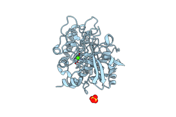 |
Organism: Alicyclobacillus sendaiensis
Method: X-RAY DIFFRACTION Resolution:2.03 Å Release Date: 2006-05-23 Classification: HYDROLASE Ligands: CA, SO4 |
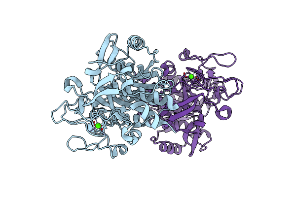 |
Organism: Alicyclobacillus sendaiensis
Method: X-RAY DIFFRACTION Resolution:2.04 Å Release Date: 2006-05-23 Classification: HYDROLASE Ligands: CA |
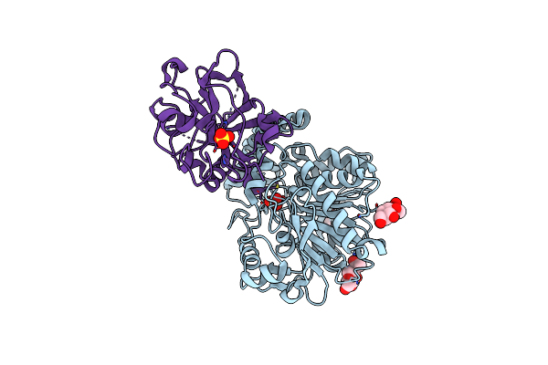 |
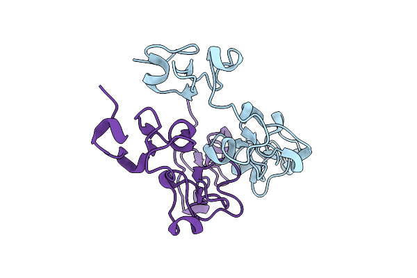
Crystal Structure Of Carboxypeptidase Y Inhibitor Complexed With The Cognate Proteinase
Organism: Saccharomyces cerevisiae
Method: X-RAY DIFFRACTION Resolution:2.70 Å Release Date: 2005-03-01 Classification: HYDROLASE Ligands: NAG, SO4 |
 |
Organism: Alicyclobacillus sendaiensis
Method: X-RAY DIFFRACTION Resolution:2.00 Å Release Date: 2004-06-01 Classification: HYDROLASE Ligands: CA |
 |
Organism: Phytolacca americana
Method: X-RAY DIFFRACTION Resolution:1.50 Å Release Date: 2004-04-13 Classification: SUGAR BINDING PROTEIN Ligands: CA |
 |
Organism: Phytolacca americana
Method: X-RAY DIFFRACTION Resolution:1.65 Å Release Date: 2004-04-13 Classification: SUGAR BINDING PROTEIN |
 |
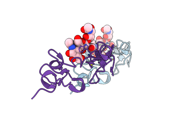
Structure Of Kumamolisin-As Complexed With A Covalently-Bound Inhibitor, Acipf
Organism: Alicyclobacillus sendaiensis
Method: X-RAY DIFFRACTION Resolution:1.80 Å Release Date: 2004-03-30 Classification: HYDROLASE/HYDROLASE INHIBITOR Ligands: SO4, CA |
 |
Organism: Alicyclobacillus sendaiensis
Method: X-RAY DIFFRACTION Resolution:2.31 Å Release Date: 2004-03-30 Classification: HYDROLASE Ligands: SO4, CA |
 |
Organism: Phytolacca americana
Method: X-RAY DIFFRACTION Resolution:1.80 Å Release Date: 2003-12-23 Classification: SUGAR BINDING PROTEIN |
 |
Crystal Structure Of Pokeweed Lectin-D2 Complexed With Tri-N-Acetylchitotriose
Organism: Phytolacca americana
Method: X-RAY DIFFRACTION Resolution:1.80 Å Release Date: 2003-12-23 Classification: SUGAR BINDING PROTEIN |
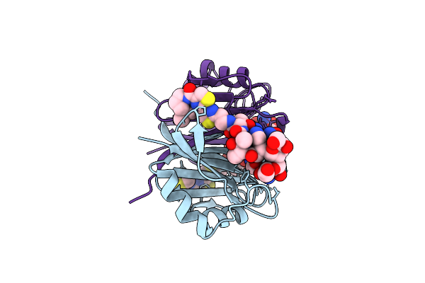 |
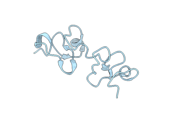
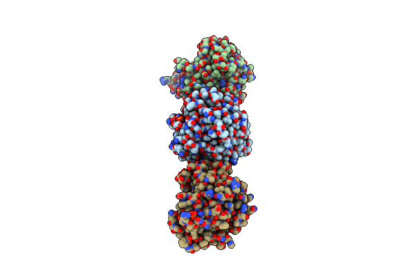
Crystal Structure Of Bleomycin-Binding Protein From Bleomycin-Producing Streptomyces Verticillus Complexed With Metal-Free Bleomycin
Organism: Streptomyces verticillus
Method: X-RAY DIFFRACTION Resolution:1.80 Å Release Date: 2002-02-06 Classification: PROTEIN BINDING Ligands: BLM |
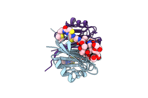 |
Crystal Structure Of Bleomycin-Binding Protein From Bleomycin-Producing Streptomyces Verticillus Complexed With Copper(Ii)-Bleomycin
Organism: Streptomyces verticillus
Method: X-RAY DIFFRACTION Resolution:1.60 Å Release Date: 2002-02-06 Classification: PROTEIN BINDING Ligands: CU, CL, BLM |
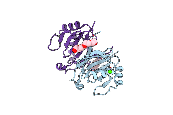 |
The 1.7 A Crystal Structure Of A Bleomycin Resistance Determinant Encoded On The Transposon Tn5
Organism: Klebsiella pneumoniae
Method: X-RAY DIFFRACTION Resolution:1.70 Å Release Date: 2001-05-02 Classification: ANTIBIOTIC INHIBITOR Ligands: CA, PG4 |
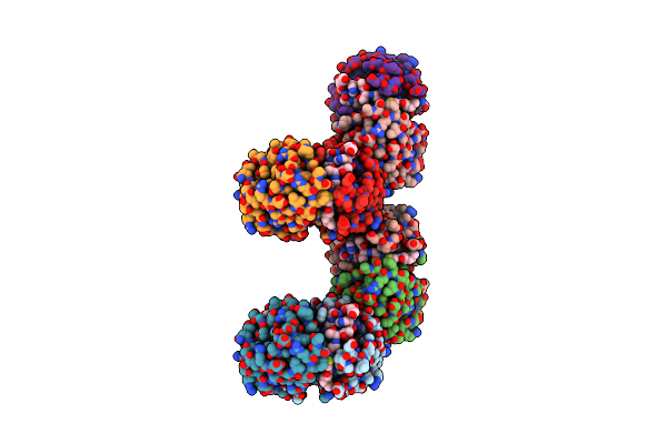 |
Organism: Klebsiella pneumoniae
Method: X-RAY DIFFRACTION Resolution:2.50 Å Release Date: 2001-04-26 Classification: ANTIBIOTIC INHIBITOR Ligands: BLM |