
Search Count: 16
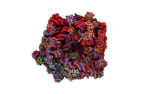 |
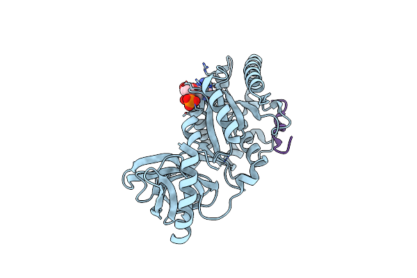
Organism: Escherichia coli
Method: ELECTRON MICROSCOPY Release Date: 2024-02-14 Classification: RIBOSOME Ligands: MG, SPD, 84D, SPM, ZN |
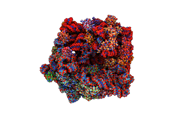 |
Organism: Escherichia coli
Method: ELECTRON MICROSCOPY Release Date: 2024-02-14 Classification: RIBOSOME Ligands: MG, SPD, 84G, SPM, ZN |
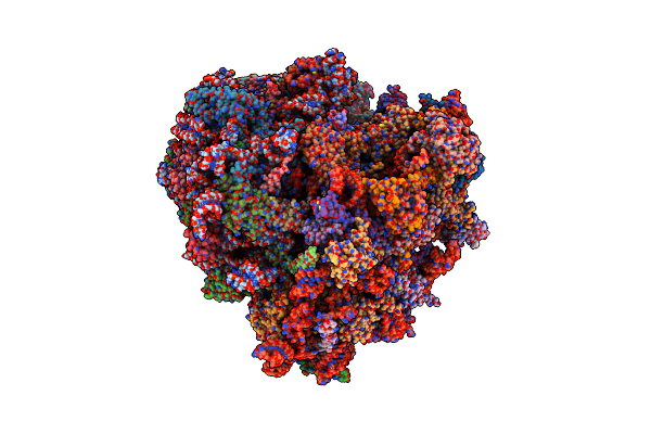 |
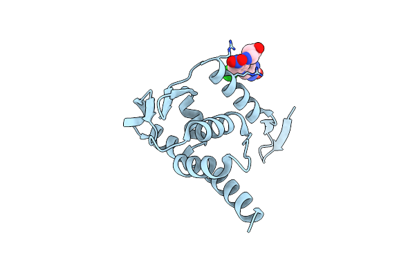
Organism: Homo sapiens
Method: ELECTRON MICROSCOPY Release Date: 2024-02-14 Classification: RIBOSOME Ligands: 84D, MG, ZN |
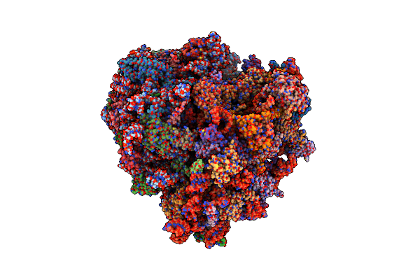 |
Organism: Homo sapiens
Method: ELECTRON MICROSCOPY Release Date: 2024-02-14 Classification: RIBOSOME Ligands: 84G, MG, ZN |
 |
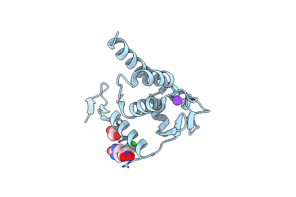
Crystal Structure Of The Complex Of Archaeal Ribosomal Stalk Protein Ap1 And Archaeal Translation Initiation Factor Aif5B
Organism: Aeropyrum pernix k1
Method: X-RAY DIFFRACTION Resolution:1.89 Å Release Date: 2018-06-27 Classification: TRANSLATION Ligands: GDP |
 |
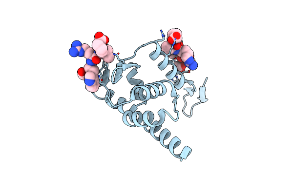
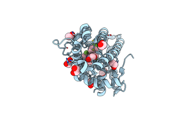
Organism: Homo sapiens
Method: X-RAY DIFFRACTION Resolution:1.58 Å Release Date: 2017-08-16 Classification: Transcription/Inhibitor Ligands: 80R, EDO, K |
 |
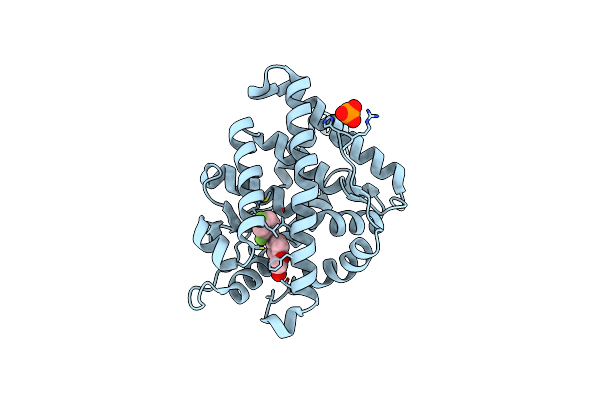
Organism: Homo sapiens
Method: X-RAY DIFFRACTION Resolution:1.86 Å Release Date: 2017-08-16 Classification: Transcription/Inhibitor Ligands: 80L |
 |
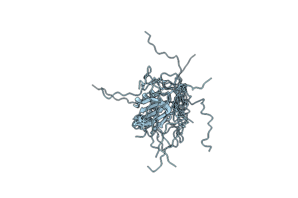
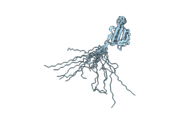
Organism: Saccharomyces cerevisiae (strain atcc 204508 / s288c)
Method: SOLUTION NMR Release Date: 2017-05-31 Classification: TRANSLATION |
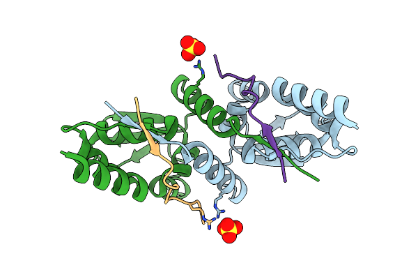 |
Organism: Homo sapiens, Enterobacteria phage t7
Method: X-RAY DIFFRACTION Resolution:1.85 Å Release Date: 2016-12-07 Classification: Transcription/Inhibitor Ligands: SO4 |
 |
Organism: Homo sapiens, Enterobacteria phage t7
Method: X-RAY DIFFRACTION Resolution:1.95 Å Release Date: 2016-12-07 Classification: Transcription/Inhibitor Ligands: EDO |
 |
Organism: Saccharomyces cerevisiae s288c
Method: SOLUTION NMR Release Date: 2016-10-26 Classification: TRANSLATION |
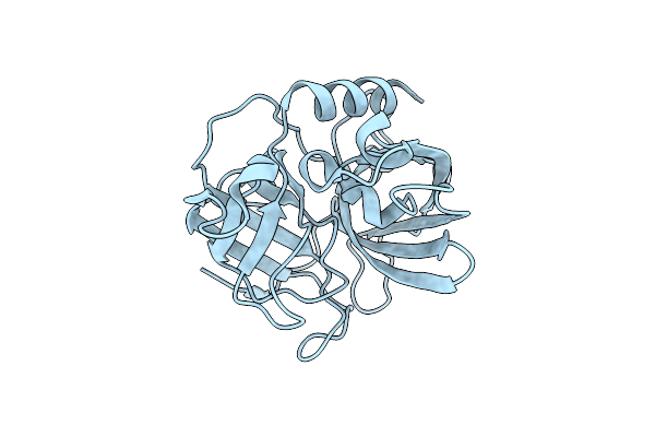 |
Organism: Arthrobacter nicotinovorans
Method: X-RAY DIFFRACTION Resolution:1.70 Å Release Date: 2015-08-26 Classification: HYDROLASE |
 |
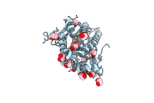
Organism: Homo sapiens
Method: X-RAY DIFFRACTION Resolution:1.10 Å Release Date: 2014-11-26 Classification: TRANSCRIPTION/TRANSCRIPTION INHIBITOR Ligands: HFN, EDO |
 |
Organism: Homo sapiens
Method: X-RAY DIFFRACTION Resolution:2.05 Å Release Date: 2013-08-21 Classification: TRANSCRIPTION/INHIBITOR Ligands: WFF, PO4 |
 |
Organism: Homo sapiens
Method: X-RAY DIFFRACTION Resolution:1.40 Å Release Date: 2013-08-21 Classification: TRANSCRIPTION/INHIBITOR Ligands: WFG, EDO |
 |
Organism: Saccharomyces cerevisiae
Method: SOLUTION NMR Release Date: 2007-11-20 Classification: TRANSLATION |