
Search Count: 89
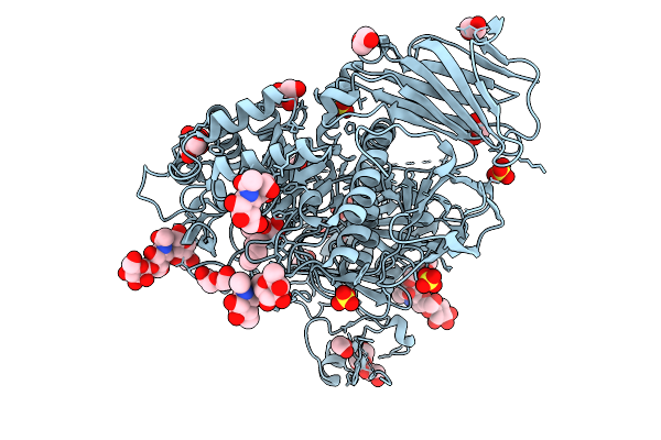 |
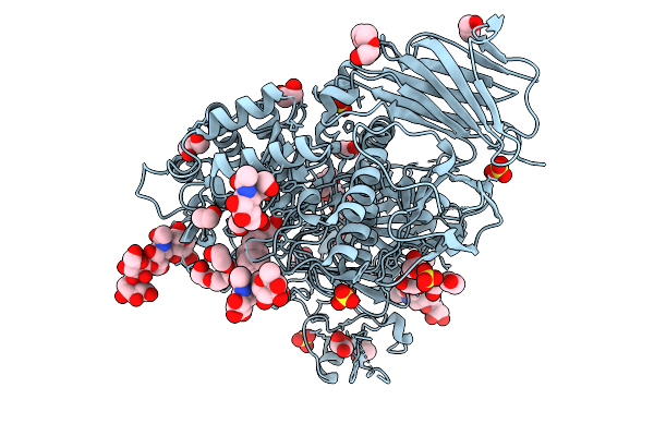
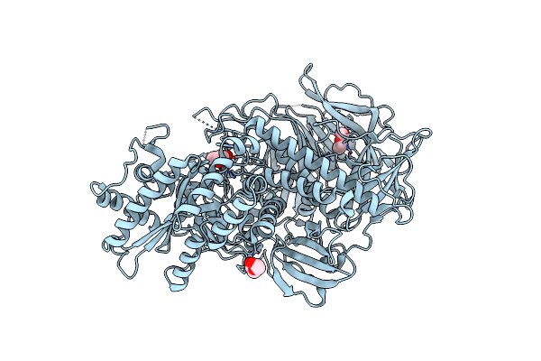
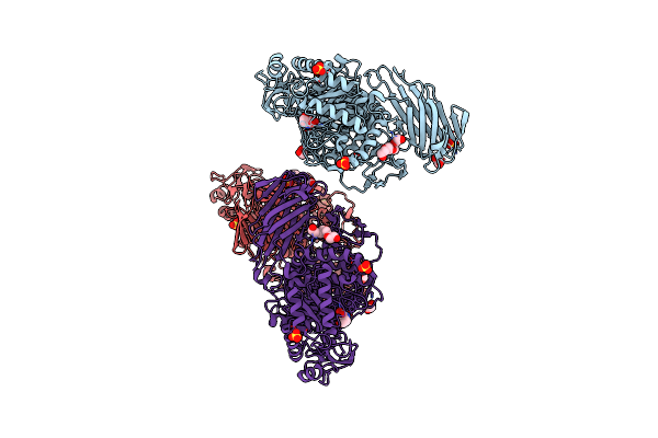
Crystal Structure Of Human Lysosomal Acid-Alpha-Glucosidase, Gaa, In Complex With Iminosugar Compound 4G
Organism: Homo sapiens
Method: X-RAY DIFFRACTION Release Date: 2025-10-01 Classification: HYDROLASE Ligands: A1IO7, SO4, CL, GOL, PEG, EDO |
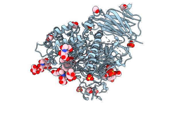 |
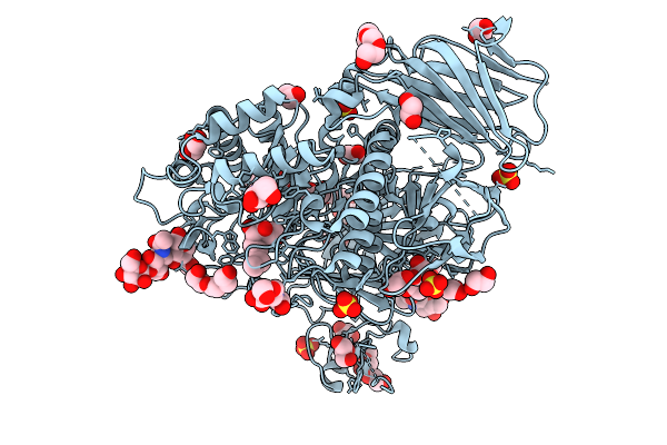
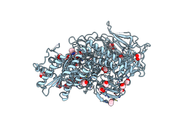
Crystal Structure Of Human Lysosomal Acid-Alpha-Glucosidase, Gaa, In Complex With Iminosugar Compound 4I
Organism: Homo sapiens
Method: X-RAY DIFFRACTION Release Date: 2025-10-01 Classification: HYDROLASE Ligands: A1IO8, SO4, CL, GOL, PEG, EDO |
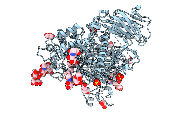 |
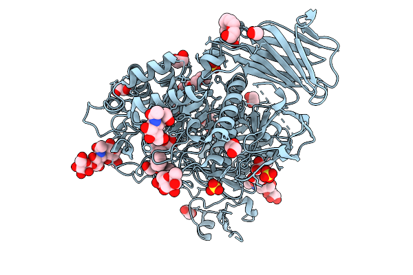
Crystal Structure Of Human Lysosomal Acid-Alpha-Glucosidase, Gaa, In Complex With Iminosugar Compound 4J
Organism: Homo sapiens
Method: X-RAY DIFFRACTION Release Date: 2025-10-01 Classification: HYDROLASE Ligands: A1IO9, SO4, CL, EDO, PEG, PGE, GOL |
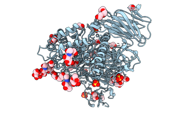 |
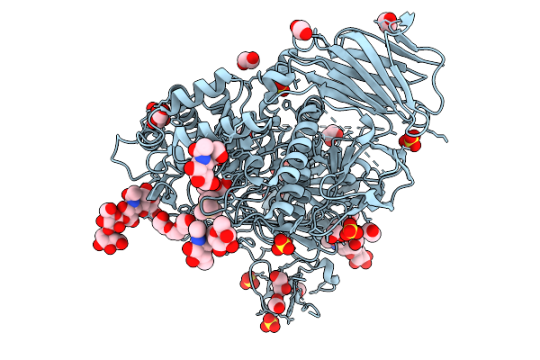
Crystal Structure Of Human Lysosomal Acid-Alpha-Glucosidase, Gaa, In Complex With Iminosugar Compound 4K
Organism: Homo sapiens
Method: X-RAY DIFFRACTION Release Date: 2025-10-01 Classification: HYDROLASE Ligands: EDO, A1IPA, SO4, CL, GOL, PGE, PEG |
 |
Crystal Structure Of Human Lysosomal Acid-Alpha-Glucosidase, Gaa, In Complex With Iminosugar Compound 4L
Organism: Homo sapiens
Method: X-RAY DIFFRACTION Release Date: 2025-10-01 Classification: HYDROLASE Ligands: A1IPB, SO4, CL, EDO, PGE, GOL, PEG |
 |
Crystal Structure Of Human Lysosomal Acid-Alpha-Glucosidase, Gaa, In Complex With Iminosugar Compound 3G
Organism: Homo sapiens
Method: X-RAY DIFFRACTION Release Date: 2025-10-01 Classification: HYDROLASE Ligands: NAG, A1IPC, SO4, CL, GOL, PGE, PEG, EDO |
 |
Crystal Structure Of Human Lysosomal Acid-Alpha-Glucosidase, Gaa, In Complex With Iminosugar Compound 4C
Organism: Homo sapiens
Method: X-RAY DIFFRACTION Release Date: 2025-09-24 Classification: HYDROLASE Ligands: A1IO6, SO4, CL, PGE, PG4, EDO |
 |
Crystal Structure Of Human Lysosomal Acid-Alpha-Glucosidase, Gaa, In Complex With Iminosugar Compound 4D
Organism: Homo sapiens
Method: X-RAY DIFFRACTION Release Date: 2025-09-24 Classification: HYDROLASE Ligands: A1IO5, SO4, CL, PEG, GOL, PGE, EDO |
 |
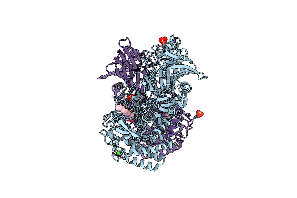
Crystal Structure Of Susa Amylase From Bacteroides Thetaiotaomicron Covalently Bound To Alpha-1,6 Branched Pseudo-Trisaccharide Activity-Based Probe
Organism: Bacteroides thetaiotaomicron
Method: X-RAY DIFFRACTION Resolution:2.43 Å Release Date: 2024-12-11 Classification: HYDROLASE Ligands: PBW, OC9, IMD, CA |
 |
Crystal Structure Of Susg From Bacteroides Thetaiotaomicron Covalently Bound To Alpha-1,6 Branched Pseudo-Trisaccharide Activity-Based Probe
Organism: Bacteroides thetaiotaomicron
Method: X-RAY DIFFRACTION Resolution:2.65 Å Release Date: 2024-12-11 Classification: HYDROLASE Ligands: PBW, OC9, A1ILG, CA, IMD, ACT |
 |
Crystal Structure Of Amylase 5 (Amy5) From Ruminococcus Bromii Covalently Bound To Alpha-1,6 Branched Pseudo-Trisaccharide Activity-Based Probe
Organism: Ruminococcus bromii
Method: X-RAY DIFFRACTION Resolution:1.40 Å Release Date: 2024-12-11 Classification: HYDROLASE Ligands: PBW, A1IHI, CA |
 |
Crystal Structure Of K38 Amylase From Bacillus Sp. Strain Ksm-K38 Covalently Bound To Alpha-1,6 Branched Pseudo-Trisaccharide Activity-Based Probe
Organism: Bacillus sp. ksm-k38
Method: X-RAY DIFFRACTION Resolution:2.02 Å Release Date: 2024-12-11 Classification: HYDROLASE Ligands: PBW, OC9, ACT, NA |
 |
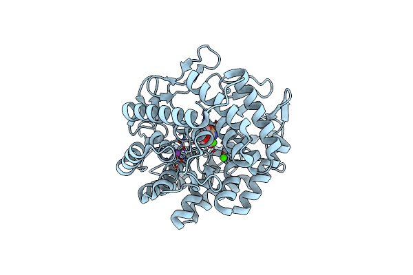
Drosophila Golgi Alpha-Mannosidase Ii (Dgmii) In Complex With Swainsonine-Configured Alkyl Indolizidine
Organism: Drosophila melanogaster
Method: X-RAY DIFFRACTION Resolution:2.47 Å Release Date: 2024-10-09 Classification: HYDROLASE Ligands: PEG, A1IGC, ZN |
 |
Drosophila Golgi Alpha-Mannosidase Ii (Dgmii) In Complex With Amide Modified Swainsonine-Configured Alkyl Indolizidine
Organism: Drosophila melanogaster
Method: X-RAY DIFFRACTION Resolution:2.14 Å Release Date: 2024-10-09 Classification: HYDROLASE Ligands: A1IGB, EDO, ZN |
 |
Organism: Thermoanaerobacterium xylanolyticum lx-11
Method: X-RAY DIFFRACTION Resolution:1.90 Å Release Date: 2024-10-02 Classification: HYDROLASE Ligands: CA, CL, SO4, XHR |
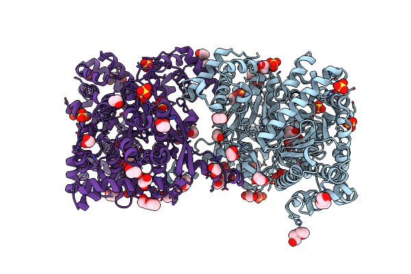 |
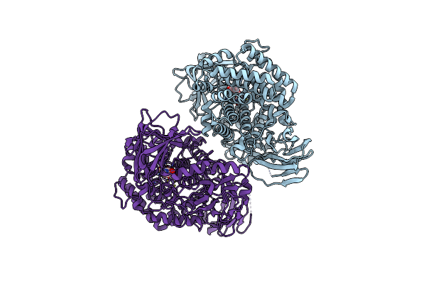
Maltodextrin Phosphorylase (Malp) In Complex With A Alpha-1,2-Cyclophellitol Analogue
Organism: Escherichia coli k-12
Method: X-RAY DIFFRACTION Resolution:2.18 Å Release Date: 2024-05-15 Classification: TRANSFERASE Ligands: A1H3V, EDO, PEG, SO4 |
 |
Crystal Structure Of Agd31B, Alpha-Transglucosylase, Complexed With A Non-Covalent 1,2- Cyclophellitol Aziridine
Organism: Cellvibrio japonicus ueda107
Method: X-RAY DIFFRACTION Resolution:1.90 Å Release Date: 2024-05-15 Classification: STRUCTURAL PROTEIN Ligands: GDQ, OXL, SO4, EDO, PG4 |
 |
Beta-L-Arabinofurano-Cyclitol Aziridines Are Cysteine-Directed Broad-Spectrum Inhibitors And Activity-Based Probes For Retaining Beta-L-Arabinofuranosidases
Organism: Bifidobacterium longum
Method: X-RAY DIFFRACTION Resolution:2.35 Å Release Date: 2023-12-13 Classification: HYDROLASE Ligands: ZN, UI5 |
 |
Gh146 Beta-L-Arabinofuranosidase From Bacteroides Thetaioatomicron In Complex With Beta-L-Arabinofurano Cyclophellitol Aziridine
Organism: Bacteroides thetaiotaomicron
Method: X-RAY DIFFRACTION Resolution:2.40 Å Release Date: 2023-12-13 Classification: HYDROLASE Ligands: UI5, ZN, UHU |
 |
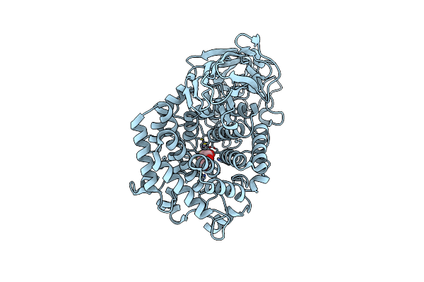
Crystal Structure Of Gh47 Alpha-1,2-Mannosidase From Caulobacter K31 Strain In Complex With Cyclosulfamidate Inhibitor
Organism: Caulobacter
Method: X-RAY DIFFRACTION Resolution:0.97 Å Release Date: 2023-10-04 Classification: HYDROLASE Ligands: 5OV, NA, CA |