
Search Count: 24
 |
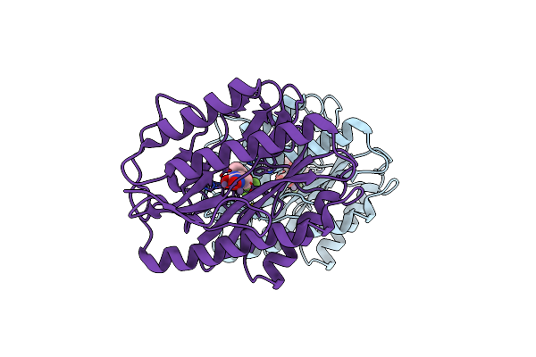
Crystal Structure Of Type 2 Pdf From Streptococcus Agalactiae, Crystallized In Imidazole Buffer
Organism: Streptococcus agalactiae
Method: X-RAY DIFFRACTION Resolution:2.00 Å Release Date: 2016-11-30 Classification: HYDROLASE Ligands: IMD, ZN |
 |
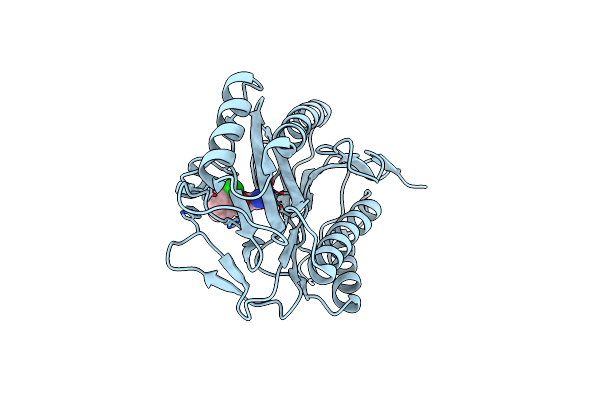
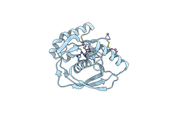
Crystal Structure Of Type 2 Pdf From Streptococcus Agalactiae, Crystallized In Cacodylate Buffer
Organism: Streptococcus agalactiae
Method: X-RAY DIFFRACTION Resolution:2.80 Å Release Date: 2016-11-30 Classification: HYDROLASE Ligands: NI |
 |
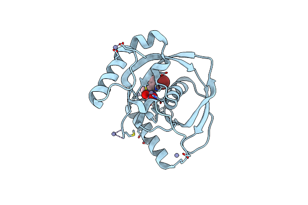
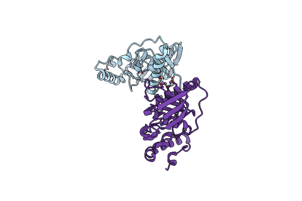
Crystal Structure Of Type 2 Pdf From Streptococcus Agalactiae In Complex With Tripeptide Met-Ala-Ser
Organism: Streptococcus agalactiae, Escherichia coli
Method: X-RAY DIFFRACTION Resolution:1.70 Å Release Date: 2016-11-30 Classification: HYDROLASE Ligands: ACT, ZN |
 |
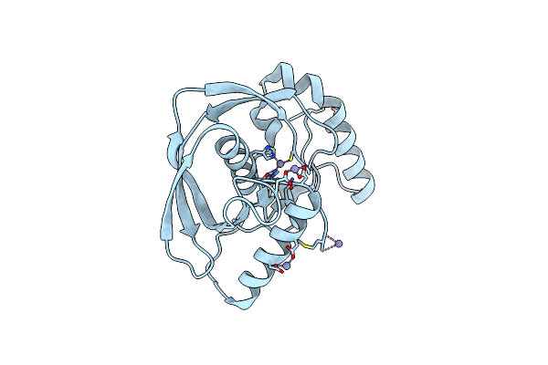
Crystal Structure Of Type 2 Pdf From Streptococcus Agalactiae In Complex With Tripeptide Met-Ala-Arg
Organism: Streptococcus agalactiae, Escherichia coli
Method: X-RAY DIFFRACTION Resolution:1.60 Å Release Date: 2016-11-30 Classification: HYDROLASE Ligands: ACT, NI |
 |
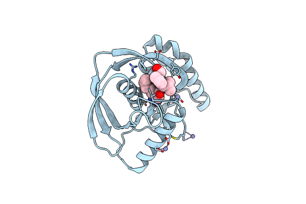
Crystal Structure Of Type 2 Pdf From Streptococcus Agalactiae In Complex With Actinonin
Organism: Streptococcus agalactiae
Method: X-RAY DIFFRACTION Resolution:2.00 Å Release Date: 2016-11-30 Classification: HYDROLASE Ligands: BB2, ACT, ZN |
 |
Crystal Structure Of Type 2 Pdf From Streptococcus Agalactiae In Complex With Inhibitor At002
Organism: Streptococcus agalactiae
Method: X-RAY DIFFRACTION Resolution:2.00 Å Release Date: 2016-11-30 Classification: HYDROLASE Ligands: SF7, ACT, IMD, ZN |
 |
Crystal Structure Of Type 2 Pdf From Streptococcus Agalactiae In Complex With Inhibitor At018
Organism: Streptococcus agalactiae
Method: X-RAY DIFFRACTION Resolution:1.60 Å Release Date: 2016-11-30 Classification: HYDROLASE Ligands: SF5, ACT, IMD, ZN |
 |
Crystal Structure Of Type 2 Pdf From Streptococcus Agalactiae In Complex With Inhibitor At019
Organism: Streptococcus agalactiae
Method: X-RAY DIFFRACTION Resolution:2.40 Å Release Date: 2016-11-30 Classification: HYDROLASE Ligands: 6JT, ACT, IMD, ZN |
 |
Crystal Structure Of Type 2 Pdf From Streptococcus Agalactiae In Complex With Inhibitor At020
Organism: Streptococcus agalactiae
Method: X-RAY DIFFRACTION Resolution:1.80 Å Release Date: 2016-11-30 Classification: HYDROLASE Ligands: 7JT, ACT, IMD, ZN |
 |
Crystal Structure Of Type 2 Pdf From Streptococcus Agalactiae In Complex With Inhibitor 6B (Ab47)
Organism: Streptococcus agalactiae
Method: X-RAY DIFFRACTION Resolution:1.70 Å Release Date: 2016-11-30 Classification: HYDROLASE Ligands: BB4, ACT, ZN |
 |
Crystal Structure Of Type 2 Pdf From Streptococcus Agalactiae In Complex With Inhibitor Smp289
Organism: Streptococcus agalactiae
Method: X-RAY DIFFRACTION Resolution:2.10 Å Release Date: 2016-11-30 Classification: HYDROLASE Ligands: 6JU, ACT, IMD, ZN |
 |
Crystal Structure Of Type 2 Pdf From Streptococcus Agalactiae In Complex With Inhibitor Ras358 (21)
Organism: Streptococcus agalactiae
Method: X-RAY DIFFRACTION Resolution:1.80 Å Release Date: 2016-11-30 Classification: HYDROLASE Ligands: PN3, ACT, IMD, ZN |
 |
Organism: Escherichia coli
Method: X-RAY DIFFRACTION Resolution:1.46 Å Release Date: 2012-06-13 Classification: HYDROLASE Ligands: MN, CO3, IKY |
 |
Organism: Escherichia coli
Method: X-RAY DIFFRACTION Resolution:1.46 Å Release Date: 2012-06-13 Classification: TRANSFERASE Ligands: MN, 5C1 |
 |
Crystal Structure Of Arabidopsis Thaliana Peptide Deformylase 1B (Atpdf1B) In Complex With Inhibitor 6B
Organism: Arabidopsis thaliana
Method: X-RAY DIFFRACTION Resolution:3.00 Å Release Date: 2011-06-08 Classification: HYDROLASE/HYDROLASE INHIBITOR Ligands: ZN, BB4 |
 |
Crystal Structure Of Arabidopsis Thaliana Petide Deformylase 1B (Atpdf1B) (Crystallized In Peg-550-Mme)
Organism: Arabidopsis thaliana
Method: X-RAY DIFFRACTION Resolution:2.00 Å Release Date: 2011-06-08 Classification: HYDROLASE Ligands: ZN |
 |
Crystal Structure Of Arabidopsis Thaliana Petide Deformylase 1B (Atpdf1B) In Complex With Inhibitor 21
Organism: Arabidopsis thaliana
Method: X-RAY DIFFRACTION Resolution:1.30 Å Release Date: 2011-06-08 Classification: Hydrolase/Hydrolase Inhibitor Ligands: PN3, ZN |
 |
Crystal Structure Of Arabidopsis Thaliana Petide Deformylase 1B (Atpdf1B) In Complex With Actinonin (Crystallized In Peg-550-Mme)
Organism: Arabidopsis thaliana
Method: X-RAY DIFFRACTION Resolution:1.90 Å Release Date: 2011-06-08 Classification: HYDROLASE/ANTIBIOTIC Ligands: BB2, ZN |
 |
Crystal Structure Of Arabidopsis Thaliana Petide Deformylase 1B (Atpdf1B) G41Q Mutant
Organism: Arabidopsis thaliana
Method: X-RAY DIFFRACTION Resolution:2.30 Å Release Date: 2011-06-08 Classification: HYDROLASE Ligands: ZN |
 |
Crystal Structure Of Arabidopsis Thaliana Petide Deformylase 1B (Atpdf1B) G41M Mutant
Organism: Arabidopsis thaliana
Method: X-RAY DIFFRACTION Resolution:2.10 Å Release Date: 2011-06-08 Classification: HYDROLASE Ligands: ZN |