
Search Count: 17
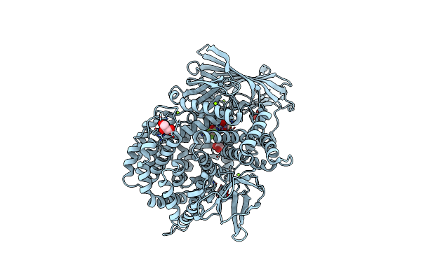 |
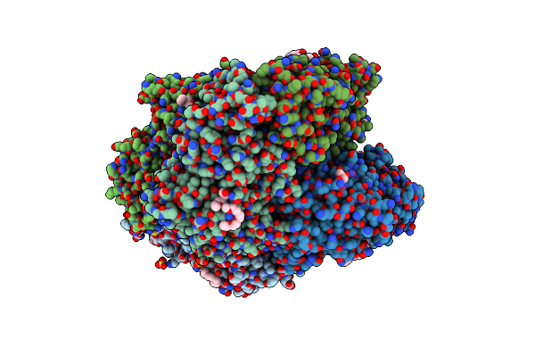
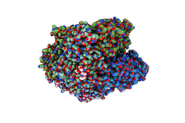
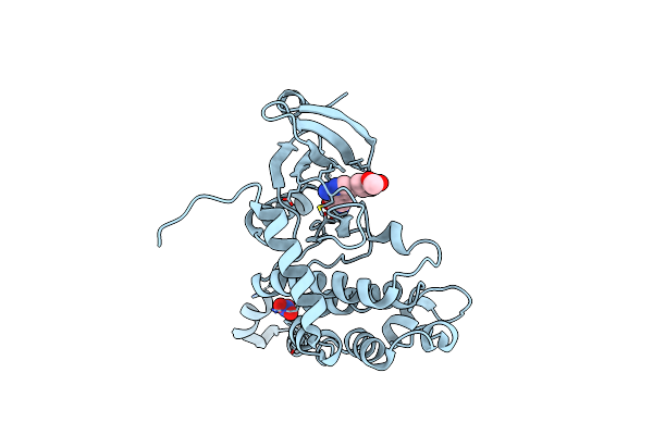
X-Ray Crystal Structure Of Pf-M1 In Complex With Inhibitor (6Da) And Catalytic Zinc Ion
Organism: Plasmodium falciparum (isolate fcb1 / columbia)
Method: X-RAY DIFFRACTION Resolution:1.82 Å Release Date: 2018-12-26 Classification: hydrolase/hydrolase inhibitor Ligands: ZN, J0Y, GOL, MG |
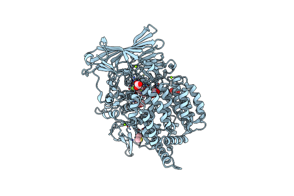 |
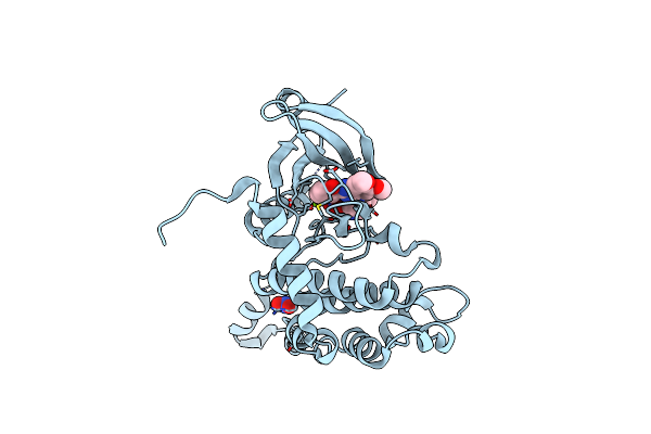
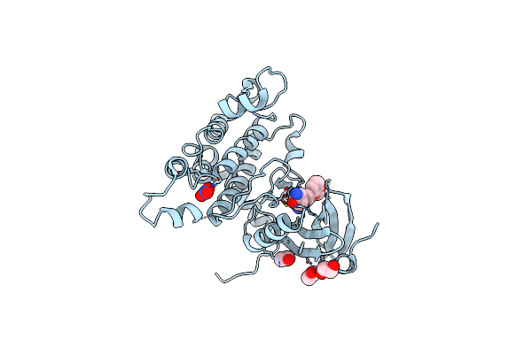
X-Ray Crystal Structure Of Pf-M1 In Complex With Inhibitor (6H) And Catalytic Zinc Ion
Organism: Plasmodium falciparum (isolate fcb1 / columbia)
Method: X-RAY DIFFRACTION Resolution:1.35 Å Release Date: 2018-12-26 Classification: hydrolase/hydrolase inhibitor Ligands: ZN, J1G, DMS, GOL, MG |
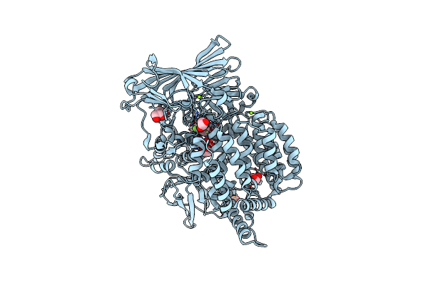 |
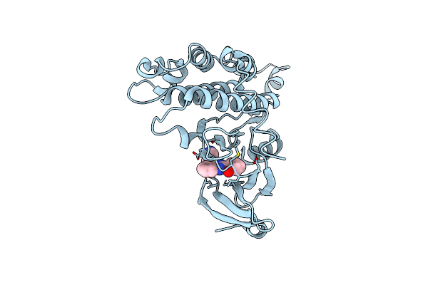
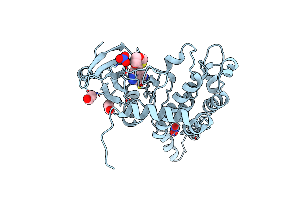
X-Ray Crystal Structure Of Pf-M1 In Complex With Inhibitor (6I) And Catalytic Zinc Ion
Organism: Plasmodium falciparum (isolate fcb1 / columbia)
Method: X-RAY DIFFRACTION Resolution:1.65 Å Release Date: 2018-12-26 Classification: hydrolase/hydrolase inhibitor Ligands: ZN, J1V, GOL, MG, PO4 |
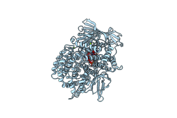 |
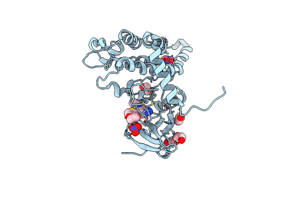
X-Ray Crystal Structure Of Pf-M1 In Complex With Inhibitor (6J) And Catalytic Zinc Ion
Organism: Plasmodium falciparum (isolate fcb1 / columbia)
Method: X-RAY DIFFRACTION Resolution:1.85 Å Release Date: 2018-12-26 Classification: hydrolase/hydrolase inhibitor Ligands: ZN, J2D, GOL, MG, PO4 |
 |
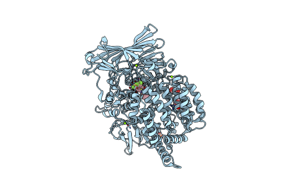
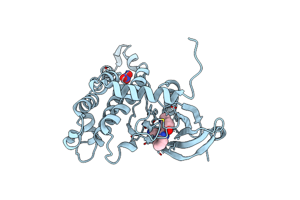
X-Ray Crystal Structure Of Pf-M17 In Complex With Inhibitor 6I And Regulatory Zinc Ion
Organism: Plasmodium falciparum nf135/5.c10
Method: X-RAY DIFFRACTION Resolution:2.10 Å Release Date: 2018-12-26 Classification: hydrolase/hydrolase inhibitor Ligands: CO3, ZN, J1V, SO4, 1PE, DMS, EDO |
 |
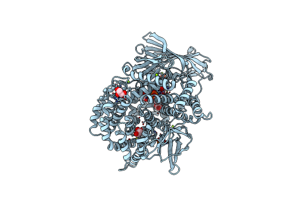
X-Ray Crystal Structure Of Pf-M1 In Complex With Inhibitor (6K) And Catalytic Zinc Ion
Organism: Plasmodium falciparum (isolate fcb1 / columbia)
Method: X-RAY DIFFRACTION Resolution:1.82 Å Release Date: 2018-12-26 Classification: hydrolase/hydrolase inhibitor Ligands: ZN, J4V, PO4, GOL, MG |
 |
X-Ray Crystal Structure Of Pf-M1 In Complex With Inhibitor (6M) And Catalytic Zinc Ion
Organism: Plasmodium falciparum (isolate fcb1 / columbia)
Method: X-RAY DIFFRACTION Resolution:1.58 Å Release Date: 2018-12-26 Classification: hydrolase/hydrolase inhibitor Ligands: ZN, J4S, GOL, PO4, MG |
 |
X-Ray Crystal Structure Of Pf-M1 In Complex With Inhibitor (6O) And Catalytic Zinc Ion
Organism: Plasmodium falciparum (isolate fcb1 / columbia)
Method: X-RAY DIFFRACTION Resolution:1.50 Å Release Date: 2018-12-26 Classification: hydrolase/hydrolase inhibitor Ligands: ZN, J4P, GOL, MG |
 |
X-Ray Crystal Structure Of Pf-M1 In Complex With Inhibitor (6P) And Catalytic Zinc Ion
Organism: Plasmodium falciparum (isolate fcb1 / columbia)
Method: X-RAY DIFFRACTION Resolution:1.50 Å Release Date: 2018-12-26 Classification: hydrolase/hydrolase inhibitor Ligands: ZN, J6A, MG, GOL, DMS, PO4 |
 |
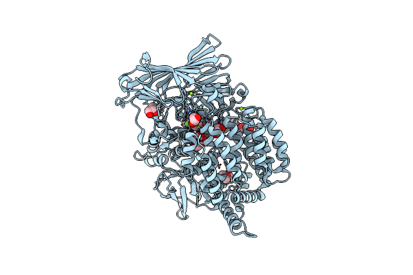
X-Ray Crystal Structure Of Pf-M17 In Complex With Inhibitor (6K) And Regulatory Zinc Ion
Organism: Plasmodium falciparum (isolate hb3)
Method: X-RAY DIFFRACTION Resolution:2.30 Å Release Date: 2018-12-26 Classification: hydrolase/hydrolase inhibitor Ligands: J4V, CO3, ZN, SO4, DMS, 1PE, EDO, 2PE |
 |
Organism: Homo sapiens
Method: X-RAY DIFFRACTION Resolution:2.30 Å Release Date: 2011-01-12 Classification: TRANSFERASE Ligands: XBJ, MG, NO3 |
 |
Co-Crystal Structure Of A Small Molecule Inhibitor Bound To The Kinase Domain Of Chk2
Organism: Homo sapiens
Method: X-RAY DIFFRACTION Resolution:3.40 Å Release Date: 2011-01-12 Classification: TRANSFERASE Ligands: B4W |
 |
Organism: Homo sapiens
Method: X-RAY DIFFRACTION Resolution:2.50 Å Release Date: 2011-01-12 Classification: TRANSFERASE Ligands: LWH, NO3 |
 |
Organism: Homo sapiens
Method: X-RAY DIFFRACTION Resolution:3.00 Å Release Date: 2009-12-29 Classification: TRANSFERASE Ligands: VGM, NO3 |
 |
Organism: Homo sapiens
Method: X-RAY DIFFRACTION Resolution:2.75 Å Release Date: 2009-12-29 Classification: TRANSFERASE Ligands: ZZK, NO3, EDO |
 |
Organism: Homo sapiens
Method: X-RAY DIFFRACTION Resolution:2.50 Å Release Date: 2009-12-29 Classification: TRANSFERASE Ligands: WTI, NO3, EDO |
 |
Organism: Homo sapiens
Method: X-RAY DIFFRACTION Resolution:2.10 Å Release Date: 2009-12-29 Classification: TRANSFERASE Ligands: WTJ, NO3, EDO |