
Search Count: 9
All
Selected
 |
Organism: Rabbit hemorrhagic disease virus
Method: ELECTRON MICROSCOPY Resolution:10.30 Å Release Date: 2012-05-23 Classification: VIRUS |
 |
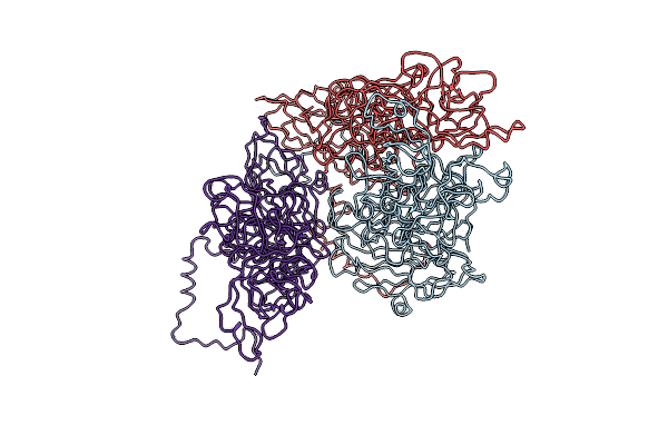
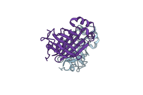
High-Resolution Structural Insights On The Sugar-Recognition And Fusion Tag Properties Of A Versatile B-Trefoil Lectin Domain
Organism: Laetiporus sulphureus
Method: X-RAY DIFFRACTION Resolution:1.47 Å Release Date: 2011-10-12 Classification: SUGAR BINDING PROTEIN |
 |
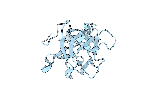
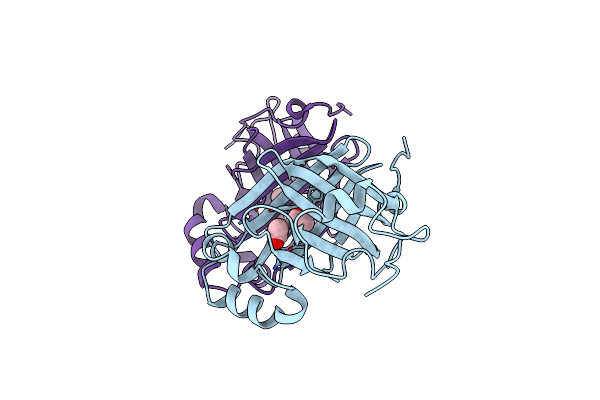
High-Resolution Structural Insights On The Sugar-Recognition And Fusion Tag Properties Of A Versatile B-Trefoil Lectin Domain
Organism: Laetiporus sulphureus
Method: X-RAY DIFFRACTION Resolution:1.67 Å Release Date: 2011-10-12 Classification: SUGAR BINDING PROTEIN Ligands: GOL |
 |
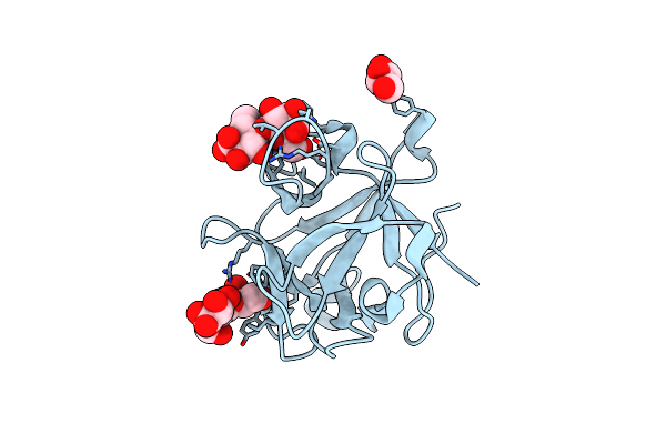
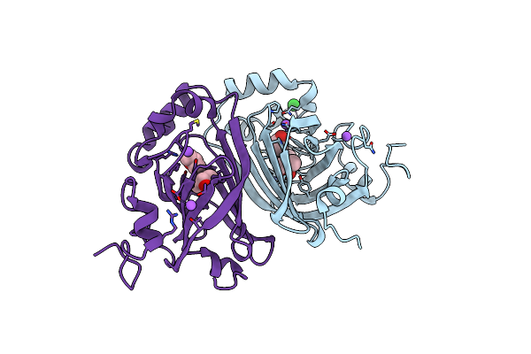
Crystal Structure Of Single Point Mutant Arg48Gln Of P-Coumaric Acid Decarboxylase From Lactobacillus Plantarum Structural Insights Into The Active Site And Decarboxylation Catalytic Mechanism
Organism: Lactobacillus plantarum
Method: X-RAY DIFFRACTION Resolution:1.73 Å Release Date: 2010-02-09 Classification: LYASE Ligands: BA |
 |
Crystal Structure Of Single Point Mutant Glu71Ser P-Coumaric Acid Decarboxylase
Organism: Lactobacillus plantarum
Method: X-RAY DIFFRACTION Resolution:2.24 Å Release Date: 2010-02-09 Classification: LYASE Ligands: BA, IPA, NA, CL |
 |
Crystal Structure Of P-Coumaric Acid Decarboxylase From Lactobacillus Plantarum: Structural Insights Into The Active Site And Decarboxylation Catalytic Mechanism
Organism: Lactobacillus plantarum
Method: X-RAY DIFFRACTION Resolution:1.38 Å Release Date: 2009-11-17 Classification: LYASE |
 |
Crystal Structure Of Single Point Mutant Tyr20Phe P-Coumaric Acid Decarboxylase From Lactobacillus Plantarum: Structural Insights Into The Active Site And Decarboxylation Catalytic Mechanism
Organism: Lactobacillus plantarum
Method: X-RAY DIFFRACTION Resolution:1.40 Å Release Date: 2009-11-17 Classification: LYASE Ligands: ACT, IPA |
 |
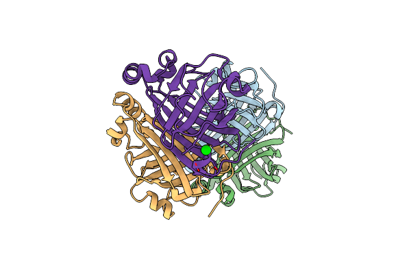
Crystal Structure Of The Hexameric Catabolic Ornithine Transcarbamylase From Lactobacillus Hilgardii
Organism: Lactobacillus hilgardii
Method: X-RAY DIFFRACTION Resolution:2.10 Å Release Date: 2009-11-17 Classification: TRANSFERASE Ligands: NI |
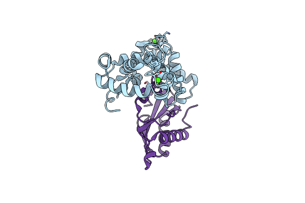 |
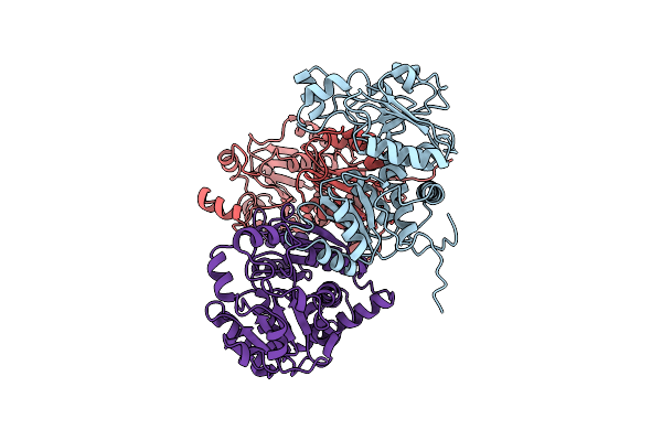
The Structure Of The C-Terminal Domain Of The Protein Kinase Atsos2 Bound To The Calcium Sensor Atsos3
Organism: Arabidopsis thaliana
Method: X-RAY DIFFRACTION Resolution:2.10 Å Release Date: 2007-09-25 Classification: SIGNALLING PROTEIN/Transferase Ligands: CA |