
Search Count: 10
 |
Organism: Streptococcus pneumoniae
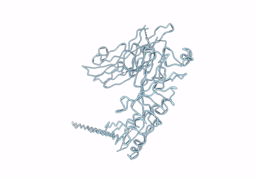
Method: X-RAY DIFFRACTION Resolution:1.98 Å Release Date: 2015-09-16 Classification: TOXIN |
 |
Organism: Streptococcus pneumoniae
Method: X-RAY DIFFRACTION Resolution:2.05 Å Release Date: 2015-09-16 Classification: TOXIN |
 |
Organism: Mus musculus
Method: X-RAY DIFFRACTION Resolution:1.70 Å Release Date: 2011-09-21 Classification: IMMUNE SYSTEM Ligands: NAG, 3TF, GOL |
 |
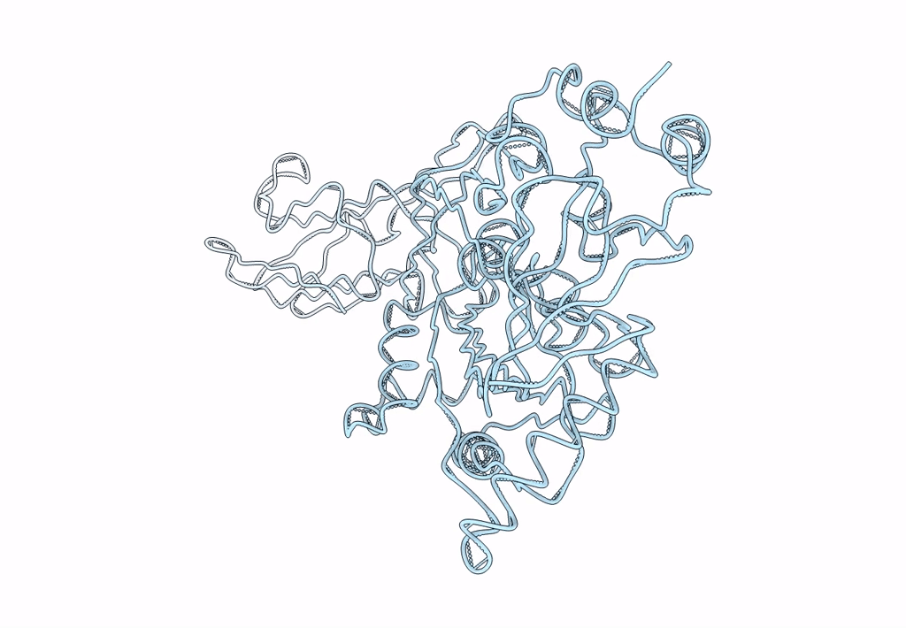
Structure Of The Catalytic Domain Of The Native Nana Sialidase From Streptococcus Pneumoniae
Organism: Streptococcus pneumoniae
Method: X-RAY DIFFRACTION Resolution:1.49 Å Release Date: 2008-12-23 Classification: HYDROLASE Ligands: MES, CL, GOL |
 |
Structure Of The Catalytic Domain Of Streptococcus Pneumoniae Sialidase Nana
Organism: Streptococcus pneumoniae
Method: X-RAY DIFFRACTION Resolution:2.50 Å Release Date: 2008-06-24 Classification: HYDROLASE Ligands: DAN, CL |
 |
Organism: Streptococcus pneumoniae
Method: X-RAY DIFFRACTION Resolution:2.30 Å Release Date: 2008-06-24 Classification: HYDROLASE Ligands: GOL |
 |
Organism: Streptococcus pneumoniae
Method: X-RAY DIFFRACTION Resolution:2.39 Å Release Date: 2008-06-24 Classification: HYDROLASE Ligands: DAN, GOL |
 |
Organism: Streptococcus pneumoniae
Method: X-RAY DIFFRACTION Resolution:1.70 Å Release Date: 2008-06-24 Classification: HYDROLASE Ligands: NHE, GOL |
 |
The Pore Structure Of Pneumolysin, Obtained By Fitting The Alpha Carbon Trace Of Perfringolysin O Into A Cryo-Em Map
Organism: Clostridium perfringens
Method: ELECTRON MICROSCOPY Resolution:29.00 Å Release Date: 2005-05-04 Classification: TOXIN |
 |
The Prepore Structure Of Pneumolysin, Obtained By Fitting The Alpha Carbon Trace Of Perfringolysin O Into A Cryo-Em Map
Organism: Clostridium perfringens
Method: ELECTRON MICROSCOPY Resolution:28.00 Å Release Date: 2005-05-04 Classification: TOXIN |