
Search Count: 48
 |
Organism: Acinetobacter silvestris
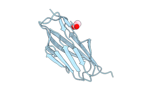
Method: X-RAY DIFFRACTION Release Date: 2025-11-26 Classification: CELL ADHESION Ligands: ACT |
 |
Organism: Drosophila melanogaster
Method: X-RAY DIFFRACTION Resolution:1.93 Å Release Date: 2025-04-02 Classification: CYTOSOLIC PROTEIN Ligands: SO4, EDO |
 |
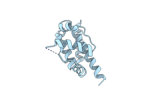
Organism: Drosophila melanogaster
Method: SOLUTION NMR Release Date: 2025-04-02 Classification: CYTOSOLIC PROTEIN |
 |
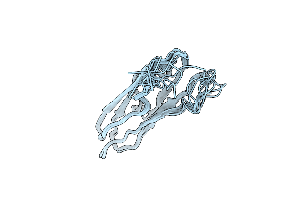
Organism: Apis dorsata
Method: X-RAY DIFFRACTION Resolution:3.50 Å Release Date: 2025-03-26 Classification: CELL CYCLE |
 |
Organism: Homo sapiens
Method: X-RAY DIFFRACTION Resolution:2.30 Å Release Date: 2022-04-06 Classification: CELL CYCLE |
 |
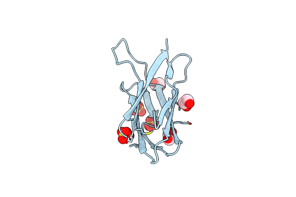
Organism: Homo sapiens
Method: X-RAY DIFFRACTION Resolution:2.08 Å Release Date: 2022-04-06 Classification: CELL CYCLE Ligands: NO3 |
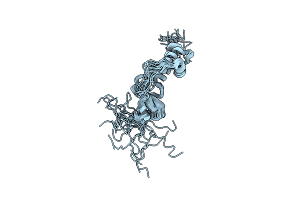 |
Organism: Homo sapiens
Method: SOLUTION NMR Release Date: 2021-09-15 Classification: PROTEIN BINDING |
 |
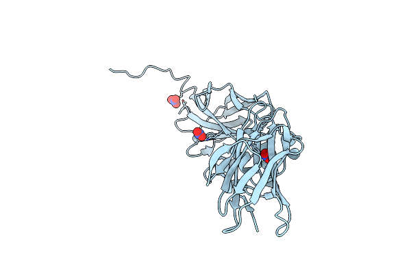
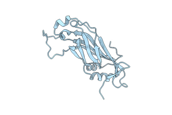
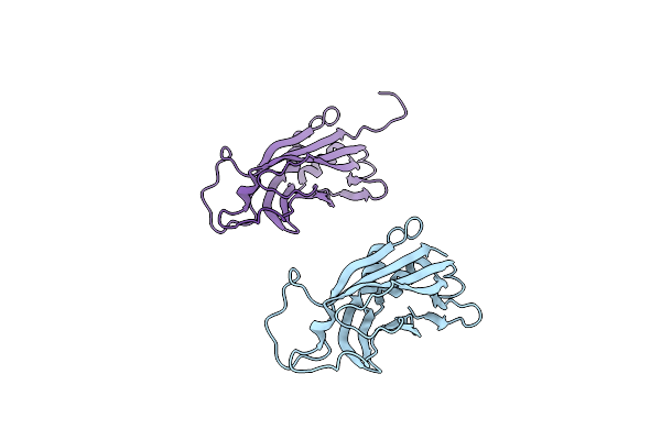
Crystal Structure Of The N-Terminal Domain Of Cep164(1-109) Bound To Camelid Nanobody 36Z
Organism: Camelidae mixed library, Homo sapiens
Method: X-RAY DIFFRACTION Resolution:1.70 Å Release Date: 2021-09-15 Classification: STRUCTURAL PROTEIN |
 |
Organism: Camelidae mixed library, Homo sapiens
Method: X-RAY DIFFRACTION Resolution:2.40 Å Release Date: 2021-09-15 Classification: STRUCTURAL PROTEIN |
 |
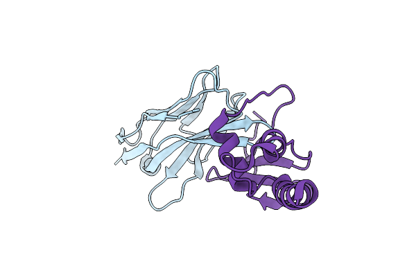
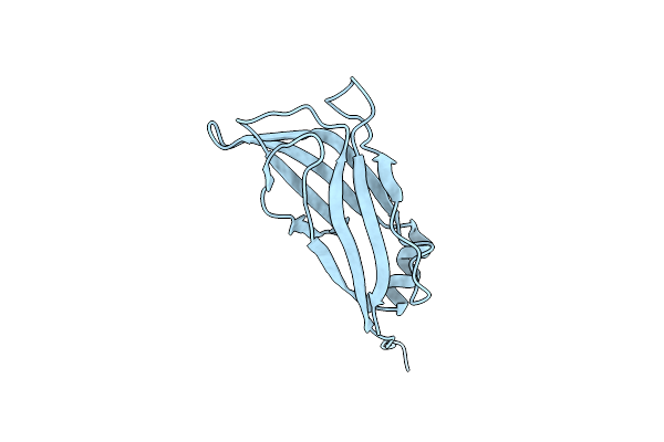
Crystal Structure Of The N-Terminal Domain Of Cep164(1-109) Bound To Camelid Nanobody 10Z
Organism: Camelidae mixed library, Homo sapiens
Method: X-RAY DIFFRACTION Resolution:1.60 Å Release Date: 2021-09-08 Classification: STRUCTURAL PROTEIN Ligands: SO4 |
 |
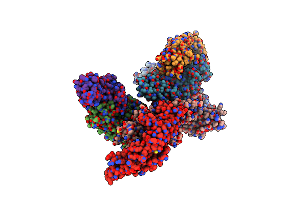
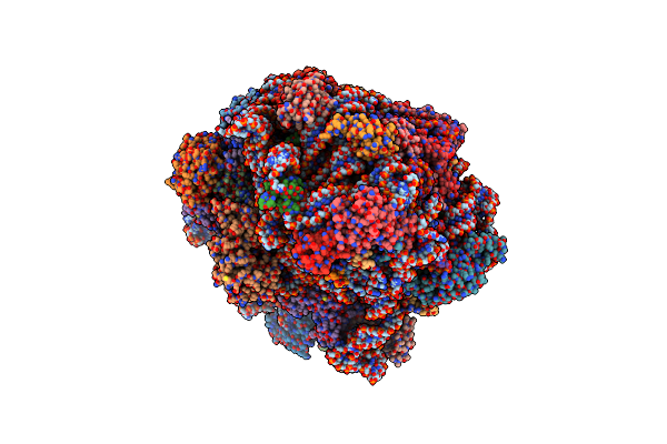
Organism: Saccharomyces cerevisiae
Method: ELECTRON MICROSCOPY Release Date: 2019-06-26 Classification: RIBOSOME Ligands: ZN |
 |
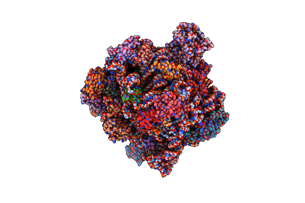
Organism: Saccharomyces cerevisiae, Saccharomyces cerevisiae
Method: ELECTRON MICROSCOPY Release Date: 2019-06-26 Classification: RIBOSOME Ligands: ZN |
 |
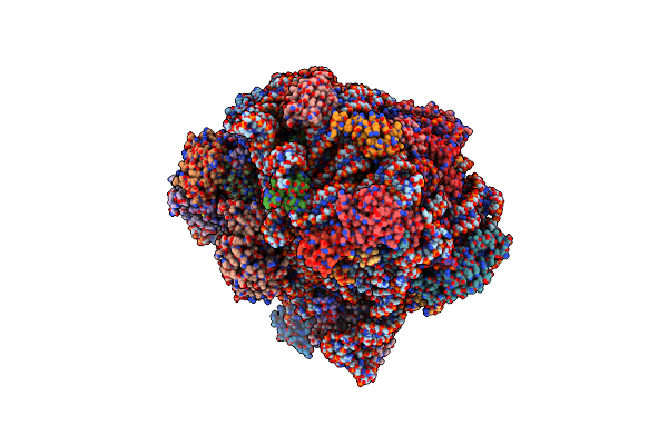
Organism: Saccharomyces cerevisiae, Saccharomyces cerevisiae
Method: ELECTRON MICROSCOPY Release Date: 2019-06-26 Classification: RIBOSOME Ligands: ZN |
 |
Organism: Saccharomyces cerevisiae
Method: ELECTRON MICROSCOPY Release Date: 2019-06-26 Classification: RIBOSOME Ligands: ZN |
 |
Organism: Saccharomyces cerevisiae
Method: ELECTRON MICROSCOPY Release Date: 2019-06-26 Classification: RIBOSOME Ligands: ZN |
 |
Organism: Saccharomyces cerevisiae
Method: ELECTRON MICROSCOPY Release Date: 2019-06-26 Classification: RIBOSOME Ligands: ZN |
 |
Organism: Oreochromis niloticus
Method: X-RAY DIFFRACTION Resolution:1.60 Å Release Date: 2018-05-02 Classification: CYTOSOLIC PROTEIN |
 |
Organism: Oreochromis niloticus
Method: X-RAY DIFFRACTION Resolution:1.50 Å Release Date: 2018-05-02 Classification: CYTOSOLIC PROTEIN |
 |
Organism: Oreochromis niloticus
Method: X-RAY DIFFRACTION Resolution:2.10 Å Release Date: 2018-05-02 Classification: CYTOSOLIC PROTEIN |
 |
Organism: Danio rerio
Method: X-RAY DIFFRACTION Resolution:1.40 Å Release Date: 2018-05-02 Classification: CYTOSOLIC PROTEIN |