
Search Count: 35
 |
Organism: Trypanosoma cruzi
Method: X-RAY DIFFRACTION Resolution:2.45 Å Release Date: 2025-02-26 Classification: UNKNOWN FUNCTION Ligands: K, GOL |
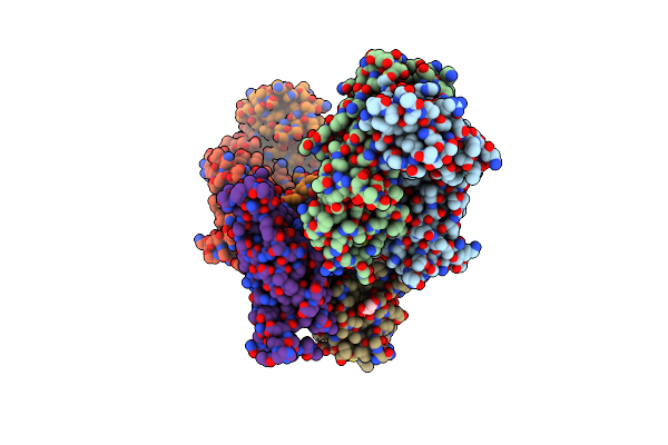 |
Organism: Homo sapiens, Severe acute respiratory syndrome coronavirus 2
Method: X-RAY DIFFRACTION Resolution:2.43 Å Release Date: 2021-05-12 Classification: IMMUNE SYSTEM/VIRAL PROTEIN Ligands: NAG |
 |
Organism: Leishmania major
Method: X-RAY DIFFRACTION Resolution:2.50 Å Release Date: 2020-02-26 Classification: DNA BINDING PROTEIN |
 |
Organism: Paenibacillus
Method: X-RAY DIFFRACTION Resolution:2.60 Å Release Date: 2018-12-05 Classification: LYASE Ligands: CA, CL, BTB, MRD, MPD |
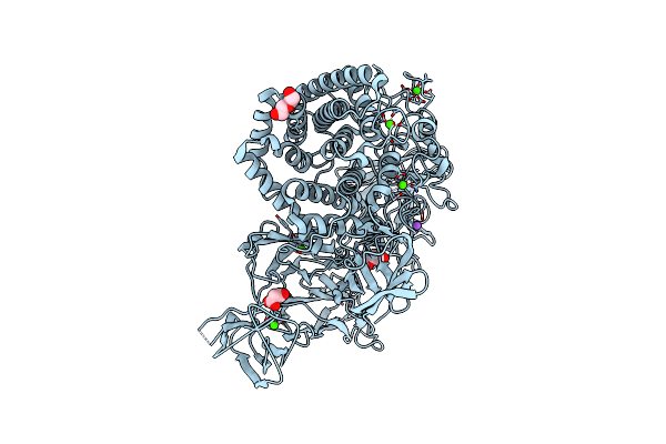 |
Structural Dynamics And Catalytic Properties Of A Multi-Modular Xanthanase, Native.
Organism: Paenibacillus
Method: X-RAY DIFFRACTION Resolution:2.04 Å Release Date: 2018-08-29 Classification: HYDROLASE Ligands: CA, NA, CL, PEG, MLI |
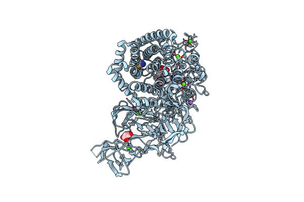 |
Structural Dynamics And Catalytic Properties Of A Multi-Modular Xanthanase (Pt Derivative)
Organism: Paenibacillus sp.
Method: X-RAY DIFFRACTION Resolution:2.00 Å Release Date: 2018-08-29 Classification: HYDROLASE Ligands: PT, B3P, SCN, CA, NA, CL, MLI |
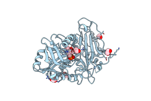 |
Single Amino Acid Variant Of Human Mitochondrial Branched Chain Amino Acid Aminotransferase 2
Organism: Homo sapiens
Method: X-RAY DIFFRACTION Resolution:1.60 Å Release Date: 2017-07-19 Classification: TRANSFERASE Ligands: PLP, EDO |
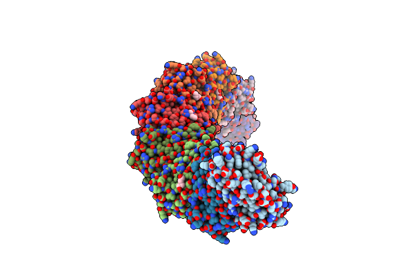 |
Organism: Sulfolobus solfataricus
Method: X-RAY DIFFRACTION Resolution:2.72 Å Release Date: 2013-02-20 Classification: IMMUNE SYSTEM Ligands: PEG |
 |
Ubiquitin Fold Modifier Conjugating Enzyme From Leishmania Major (Probable)
Organism: Leishmania major
Method: X-RAY DIFFRACTION Resolution:2.20 Å Release Date: 2009-12-01 Classification: LIGASE |
 |
Organism: Leishmania major
Method: X-RAY DIFFRACTION Resolution:1.66 Å Release Date: 2008-09-09 Classification: OXIDOREDUCTASE Ligands: ACT, 0PA |
 |
Organism: Leishmania major
Method: X-RAY DIFFRACTION Resolution:2.50 Å Release Date: 2008-09-09 Classification: OXIDOREDUCTASE Ligands: ACT, FIC |
 |
Organism: Mytilus edulis
Method: X-RAY DIFFRACTION Resolution:1.60 Å Release Date: 2006-02-22 Classification: HYDROLASE Ligands: SO4 |
 |
Organism: Plasmodium falciparum
Method: X-RAY DIFFRACTION Resolution:2.09 Å Release Date: 2005-12-27 Classification: ISOMERASE Ligands: PO4 |
 |
Crystal Structure Of Nucleoside 2-Deoxyribosyltransferase From Trypanosoma Brucei At 1.5 A Resolution With (2-Ethylphenyl)Methanol Bound
Organism: Trypanosoma brucei
Method: X-RAY DIFFRACTION Resolution:1.50 Å Release Date: 2005-12-06 Classification: TRANSFERASE Ligands: SO4, 12M, GOL |
 |
Crystal Structure Of Nucleoside 2-Deoxyribosyltransferase From Trypanosoma Brucei At 1.6 A Resolution With 1-Methylquinolin-2(1H)-One Bound
Organism: Trypanosoma brucei
Method: X-RAY DIFFRACTION Resolution:1.60 Å Release Date: 2005-12-06 Classification: TRANSFERASE Ligands: SO4, 12Q, GOL |
 |
Crystal Structure Of Nucleoside 2-Deoxyribosyltransferase From Trypanosoma Brucei At 1.6 A Resolution With Benzo[Cd]Indol-2(1H)-One Bound
Organism: Trypanosoma brucei
Method: X-RAY DIFFRACTION Resolution:1.60 Å Release Date: 2005-12-06 Classification: TRANSFERASE Ligands: SO4, 12B, GOL |
 |
Crystal Structure Of Nucleoside 2-Deoxyribosyltransferase From Trypanosoma Brucei At 1.7 A Resolution With 5-Aminoisoquinoline Bound
Organism: Trypanosoma brucei
Method: X-RAY DIFFRACTION Resolution:1.70 Å Release Date: 2005-11-22 Classification: TRANSFERASE Ligands: SO4, 5IQ, GOL |
 |
Crystal Structure Of Glyceraldehyde-3-Phosphate Dehydrogenase From Plasmodium Falciparum At 2.25 Angstrom Resolution Reveals Intriguing Extra Electron Density In The Active Site
Organism: Plasmodium falciparum
Method: X-RAY DIFFRACTION Resolution:2.25 Å Release Date: 2005-10-04 Classification: OXIDOREDUCTASE Ligands: NAD, AES, GOL |
 |
Crystal Structure Of Glyceraldehyde-3-Phosphate Dehydrogenase From Plasmodium Falciparum At 2.25 Angstrom Resolution Reveals Intriguing Extra Electron Density In The Active Site
Organism: Plasmodium falciparum
Method: X-RAY DIFFRACTION Resolution:2.50 Å Release Date: 2005-10-04 Classification: OXIDOREDUCTASE Ligands: NAD, AES |
 |
The Structure Of A Modular Endo-Beta-1,4-Mannanase From Cellulomonas Fimi Explains The Product Specificity Of Glycoside Hydrolase Family 26 Mannanases.
Organism: Cellulomonas fimi
Method: X-RAY DIFFRACTION Resolution:2.90 Å Release Date: 2005-09-26 Classification: HYDROLASE Ligands: CAC |