
Search Count: 26
 |
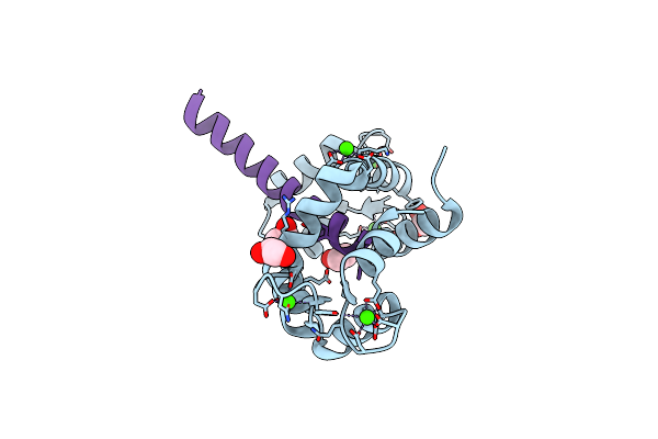
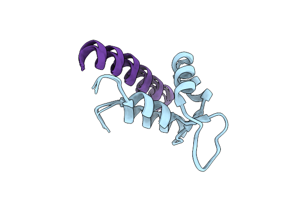
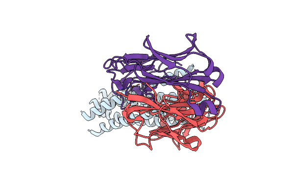
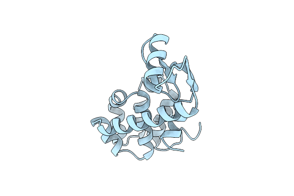
Nmr Structure Of Half-Calcified Calmodulin Mutant (Camef12) Bound To The Iq-Motif Of Cav1.2
Organism: Homo sapiens
Method: SOLUTION NMR Release Date: 2022-07-06 Classification: METAL BINDING PROTEIN Ligands: CA |
 |
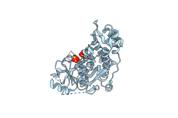
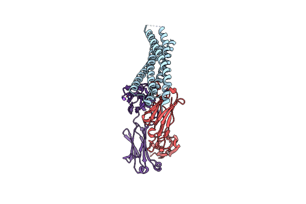
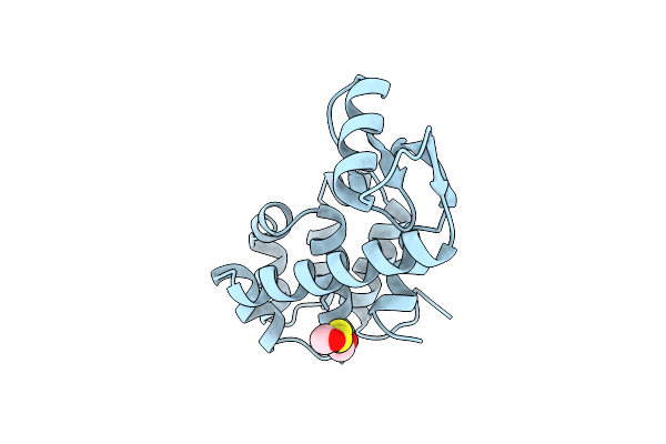
Structure Of Calmodulin Bound To The Cardiac Ryanodine Receptor (Ryr2) At Residues: Phe4246 To Val4271
Organism: Homo sapiens
Method: X-RAY DIFFRACTION Resolution:1.65 Å Release Date: 2021-04-07 Classification: METAL BINDING PROTEIN Ligands: CA, EDO |
 |
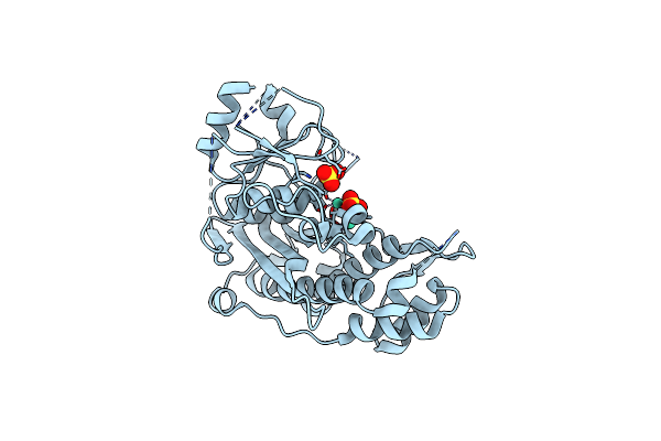
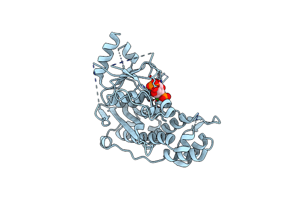
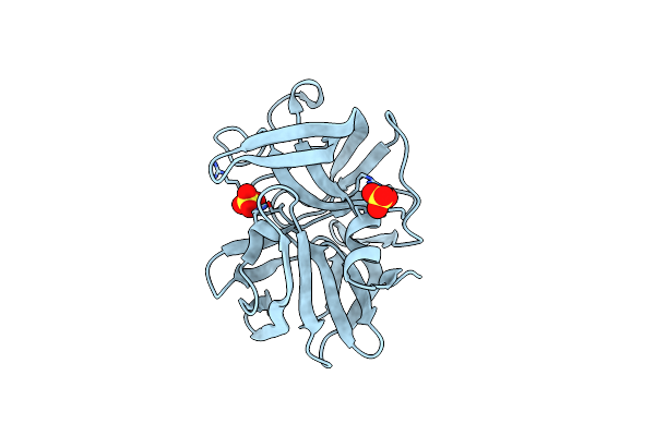
Crystal Structure Of Inositol Polyphosphate 1-Phosphatase Inpp1 In Complex Gadolinium After Addition Of Inositol 1,3,4-Trisphosphate At 2.5 Angstrom Resolution
Organism: Bos taurus
Method: X-RAY DIFFRACTION Resolution:2.80 Å Release Date: 2020-11-25 Classification: SIGNALING PROTEIN Ligands: SO4, GD |
 |
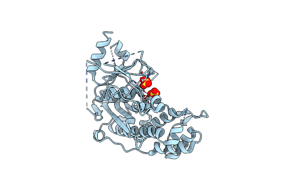
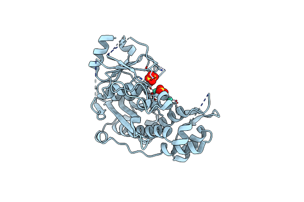
Crystal Structure Of Inositol Polyphosphate 1-Phosphatase Inpp1 In Complex Gadolinium After Addition Of Inositol 1,3,4-Trisphosphate And Lithium At 3.2 Angstrom Resolution
Organism: Bos taurus
Method: X-RAY DIFFRACTION Resolution:3.20 Å Release Date: 2020-11-25 Classification: SIGNALING PROTEIN Ligands: GD, SO4 |
 |
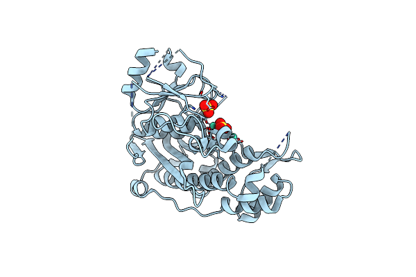
Crystal Structure Of Inositol Polyphosphate 1-Phosphatase (Inpp1) D54A Mutant
Organism: Bos taurus
Method: X-RAY DIFFRACTION Resolution:2.40 Å Release Date: 2020-11-25 Classification: HYDROLASE Ligands: CA, SO4 |
 |
Crystal Structure Of Inositol Polyphosphate 1-Phosphatase (Inpp1) D54A Mutant In Complex With Inositol (1,4)-Bisphosphate
Organism: Bos taurus
Method: X-RAY DIFFRACTION Resolution:2.60 Å Release Date: 2020-11-25 Classification: HYDROLASE Ligands: 2IP, CA |
 |
Crystal Structure Of Inositol Polyphosphate 1-Phosphatase Inpp1 In Complex Gadolinium But No Lithium At 3 Angstrom Resolution
Organism: Bos taurus
Method: X-RAY DIFFRACTION Resolution:3.00 Å Release Date: 2020-11-18 Classification: SIGNALING PROTEIN Ligands: GD, SO4 |
 |
Crystal Structure Of Inositol Polyphosphate 1-Phosphatase Inpp1 In Complex Gadolinium And Lithium At 2.5 Angstrom Resolution
Organism: Bos taurus
Method: X-RAY DIFFRACTION Resolution:2.50 Å Release Date: 2020-11-18 Classification: SIGNALING PROTEIN Ligands: GD, SO4 |
 |
Organism: Xenopus laevis, Oryctolagus cuniculus
Method: SOLUTION NMR Release Date: 2019-09-25 Classification: PROTEIN BINDING |
 |
Organism: Mus musculus, Homo sapiens
Method: SOLUTION NMR Release Date: 2019-01-16 Classification: SIGNALING PROTEIN |
 |
Organism: Plasmodium falciparum 3d7
Method: SOLUTION NMR Release Date: 2012-04-18 Classification: MEMBRANE PROTEIN |
 |
Organism: Human immunodeficiency virus 1, Homo sapiens
Method: X-RAY DIFFRACTION Resolution:2.50 Å Release Date: 2010-12-08 Classification: IMMUNE SYSTEM |
 |
Organism: Human immunodeficiency virus 1, Homo sapiens
Method: X-RAY DIFFRACTION Resolution:2.05 Å Release Date: 2010-12-01 Classification: IMMUNE SYSTEM |
 |
Crystal Structure Of Yopm-Leucine Rich Effector Protein From Yersinia Pestis
Organism: Yersinia pestis
Method: X-RAY DIFFRACTION Resolution:2.35 Å Release Date: 2001-10-10 Classification: TOXIN Ligands: ACT, CA, HG |
 |
Novel Molecular Architecture Of Yopm-A Leucine-Rich Effector Protein From Yersinia Pestis
Organism: Yersinia pestis
Method: X-RAY DIFFRACTION Resolution:2.10 Å Release Date: 2001-10-10 Classification: TOXIN Ligands: CA |
 |
The Crystal Structure Of Liganded Maltodextrin-Binding Protein From Pyrococcus Furiosus
Organism: Pyrococcus furiosus
Method: X-RAY DIFFRACTION Resolution:1.85 Å Release Date: 2001-01-19 Classification: SUGAR BINDING PROTEIN Ligands: SO4 |
 |
Pro Region C-Terminus: Protease Active Site Interactions Are Critical In Catalyzing The Folding Of Alpha-Lytic Protease
Organism: Lysobacter enzymogenes
Method: X-RAY DIFFRACTION Resolution:2.10 Å Release Date: 1998-08-12 Classification: HYDROLASE Ligands: SO4 |
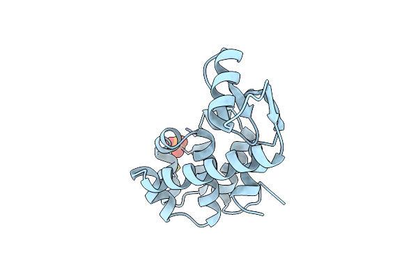 |
Hydrophobic Core Repacking And Aromatic-Aromatic Interaction In The Thermostable Mutant Of T4 Lysozyme Ser 117 (Right Arrow) Phe
Organism: Enterobacteria phage t4
Method: X-RAY DIFFRACTION Resolution:2.00 Å Release Date: 1993-07-15 Classification: HYDROLASE (O-GLYCOSYL) Ligands: PO4, CL |
 |
The Structural And Thermodynamic Consequences Of Burying A Charged Residue Within The Hydrophobic Core Of T4 Lysozyme
Organism: Enterobacteria phage t4
Method: X-RAY DIFFRACTION Resolution:1.90 Å Release Date: 1991-10-15 Classification: HYDROLASE (O-GLYCOSYL) |
 |
Analysis Of The Interaction Between Charged Side Chains And The Alpha-Helix Dipole Using Designed Thermostable Mutants Of Phage T4 Lysozyme
Organism: Enterobacteria phage t4
Method: X-RAY DIFFRACTION Resolution:1.90 Å Release Date: 1991-10-15 Classification: HYDROLASE (O-GLYCOSYL) Ligands: CL, BME |