
Search Count: 132
 |
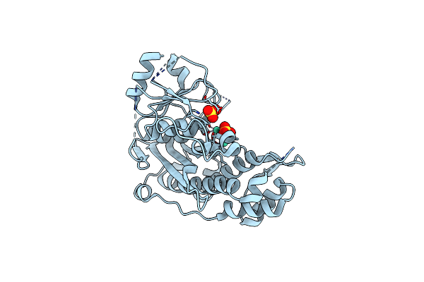
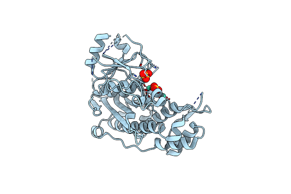
Crystal Structure Of Sars-Cov-2 Main Protease (Mpro)In Complex With Inhibitor Avi-3318
Organism: Severe acute respiratory syndrome coronavirus 2
Method: X-RAY DIFFRACTION Release Date: 2025-07-23 Classification: HYDROLASE/HYDROLASE INHIBITOR Ligands: A1BTM |
 |
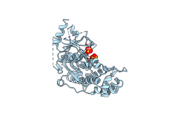
Crystal Structure Of Sars-Cov-2 Main Protease (Mpro) In Complex With Inhibitor Avi-4692
Organism: Severe acute respiratory syndrome coronavirus 2
Method: X-RAY DIFFRACTION Release Date: 2025-07-23 Classification: HYDROLASE Ligands: A1BVT, A1BTO |
 |
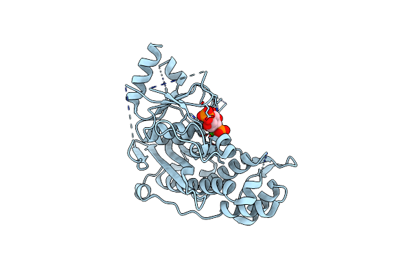
Crystal Structure Of Sars-Cov-2 Main Protease (Mpro)In Complex With Inhibitor Avi-4516
Organism: Severe acute respiratory syndrome coronavirus 2
Method: X-RAY DIFFRACTION Release Date: 2025-07-23 Classification: HYDROLASE/HYDROLASE INHIBITOR Ligands: A1BTP, EDO |
 |
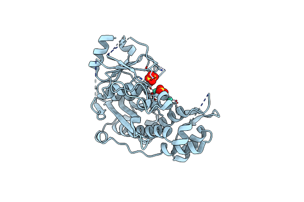
Crystal Structure Of Sars-Cov-2 Main Protease (Mpro) Variant Q192T In Complex With Inhibitor Avi-4303
Organism: Severe acute respiratory syndrome coronavirus 2
Method: X-RAY DIFFRACTION Release Date: 2025-07-23 Classification: HYDROLASE/HYDROLASE INHIBITOR Ligands: EDO, A1BTN, CL |
 |
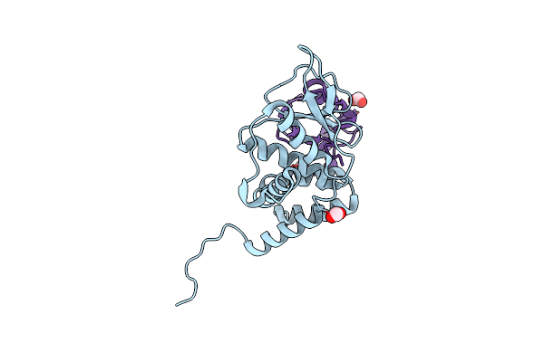
Nmr Structure Of Half-Calcified Calmodulin Mutant (Camef12) Bound To The Iq-Motif Of Cav1.2
Organism: Homo sapiens
Method: SOLUTION NMR Release Date: 2022-07-06 Classification: METAL BINDING PROTEIN Ligands: CA |
 |
Structure Of Calmodulin Bound To The Cardiac Ryanodine Receptor (Ryr2) At Residues: Phe4246 To Val4271
Organism: Homo sapiens
Method: X-RAY DIFFRACTION Resolution:1.65 Å Release Date: 2021-04-07 Classification: METAL BINDING PROTEIN Ligands: CA, EDO |
 |
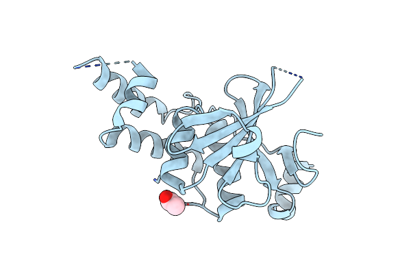
Aalall Segment From The Nucleoprotein Of Sars-Cov-2, Residues 217-222, Crystal Form 1
Organism: Severe acute respiratory syndrome coronavirus 2
Method: X-RAY DIFFRACTION Resolution:1.12 Å Release Date: 2021-03-17 Classification: PROTEIN FIBRIL Ligands: TFA |
 |
Aalall Segment From The Nucleoprotein Of Sars-Cov-2, Residues 217-222, Crystal Form 2
Organism: Severe acute respiratory syndrome coronavirus 2
Method: X-RAY DIFFRACTION Resolution:1.30 Å Release Date: 2021-03-17 Classification: PROTEIN FIBRIL Ligands: PG4 |
 |
Organism: Severe acute respiratory syndrome coronavirus 2
Method: X-RAY DIFFRACTION Resolution:1.10 Å Release Date: 2021-03-17 Classification: PROTEIN FIBRIL |
 |
Organism: Severe acute respiratory syndrome coronavirus 2
Method: X-RAY DIFFRACTION Resolution:1.30 Å Release Date: 2021-03-17 Classification: PROTEIN FIBRIL |
 |
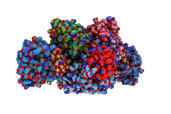
Crystal Structure Of Inositol Polyphosphate 1-Phosphatase Inpp1 In Complex Gadolinium After Addition Of Inositol 1,3,4-Trisphosphate At 2.5 Angstrom Resolution
Organism: Bos taurus
Method: X-RAY DIFFRACTION Resolution:2.80 Å Release Date: 2020-11-25 Classification: SIGNALING PROTEIN Ligands: SO4, GD |
 |
Crystal Structure Of Inositol Polyphosphate 1-Phosphatase Inpp1 In Complex Gadolinium After Addition Of Inositol 1,3,4-Trisphosphate And Lithium At 3.2 Angstrom Resolution
Organism: Bos taurus
Method: X-RAY DIFFRACTION Resolution:3.20 Å Release Date: 2020-11-25 Classification: SIGNALING PROTEIN Ligands: GD, SO4 |
 |
Crystal Structure Of Inositol Polyphosphate 1-Phosphatase (Inpp1) D54A Mutant
Organism: Bos taurus
Method: X-RAY DIFFRACTION Resolution:2.40 Å Release Date: 2020-11-25 Classification: HYDROLASE Ligands: CA, SO4 |
 |
Crystal Structure Of Inositol Polyphosphate 1-Phosphatase (Inpp1) D54A Mutant In Complex With Inositol (1,4)-Bisphosphate
Organism: Bos taurus
Method: X-RAY DIFFRACTION Resolution:2.60 Å Release Date: 2020-11-25 Classification: HYDROLASE Ligands: 2IP, CA |
 |
Crystal Structure Of Inositol Polyphosphate 1-Phosphatase Inpp1 In Complex Gadolinium But No Lithium At 3 Angstrom Resolution
Organism: Bos taurus
Method: X-RAY DIFFRACTION Resolution:3.00 Å Release Date: 2020-11-18 Classification: SIGNALING PROTEIN Ligands: GD, SO4 |
 |
Crystal Structure Of Inositol Polyphosphate 1-Phosphatase Inpp1 In Complex Gadolinium And Lithium At 2.5 Angstrom Resolution
Organism: Bos taurus
Method: X-RAY DIFFRACTION Resolution:2.50 Å Release Date: 2020-11-18 Classification: SIGNALING PROTEIN Ligands: GD, SO4 |
 |
Organism: Wolbachia pipientis wmel
Method: X-RAY DIFFRACTION Resolution:1.47 Å Release Date: 2020-07-01 Classification: HYDROLASE Ligands: ACT |
 |
Crystal Structure Of An Otu Deubiquitinase From Wolbachia Pipientis Wmel Bound To Ubiquitin
Organism: Homo sapiens, Wolbachia pipientis wmel
Method: X-RAY DIFFRACTION Resolution:1.82 Å Release Date: 2020-07-01 Classification: HYDROLASE Ligands: FLC |
 |
Crystal Structure Of An Otu Deubiquitinase From Escherichia Albertii Bound To Ubiquitin
Organism: Escherichia albertii (strain tw07627), Homo sapiens
Method: X-RAY DIFFRACTION Resolution:2.10 Å Release Date: 2020-07-01 Classification: HYDROLASE Ligands: FMT |
 |
Organism: Clostridioides difficile
Method: ELECTRON MICROSCOPY Release Date: 2020-03-18 Classification: TRANSLOCASE Ligands: CA |