Search Count: 48
 |
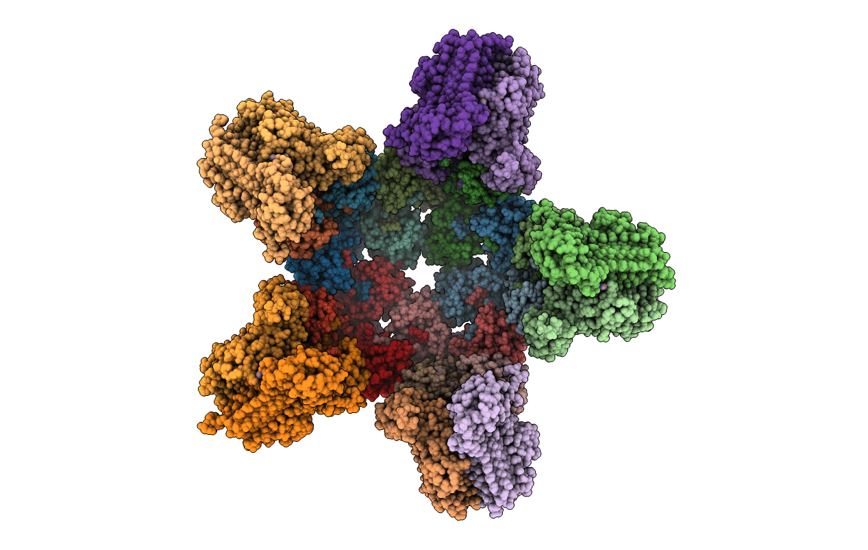
Crystal Structure Of Pseudomonas Aeruginosa Lpxa In Complex With Compound 1
Organism: Pseudomonas aeruginosa (strain pa7)
Method: X-RAY DIFFRACTION Resolution:1.84 Å Release Date: 2021-10-13 Classification: TRANSFERASE Ligands: VFE, CL, ACT |
 |
Crystal Structure Of Pseudomonas Aeruginosa Lpxa In Complex With Compound 1
Organism: Pseudomonas aeruginosa (strain pa7)
Method: X-RAY DIFFRACTION Resolution:2.84 Å Release Date: 2021-10-13 Classification: TRANSFERASE Ligands: VFE, MG |
 |
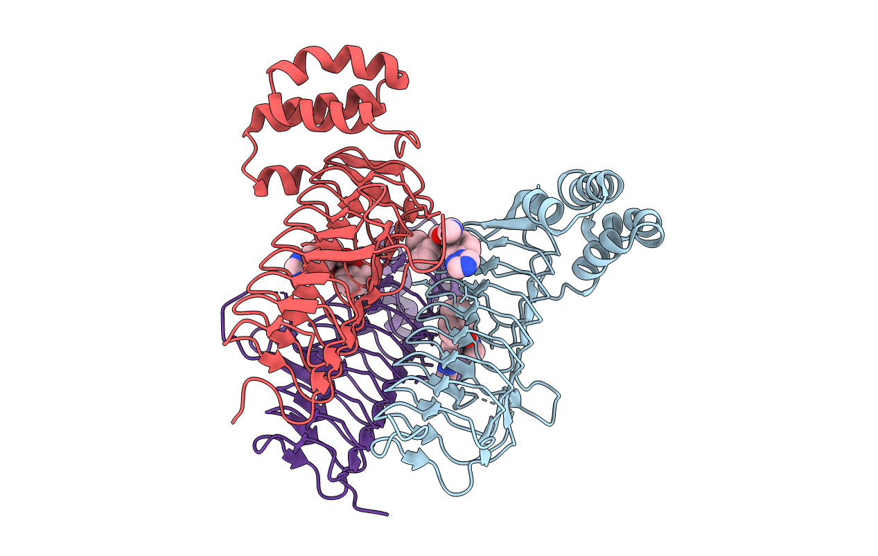
Crystal Structure Of Pseudomonas Aeruginosa Lpxa In Complex With Compound 7
Organism: Pseudomonas aeruginosa (strain pa7)
Method: X-RAY DIFFRACTION Resolution:1.87 Å Release Date: 2021-10-13 Classification: TRANSFERASE Ligands: VJE |
 |
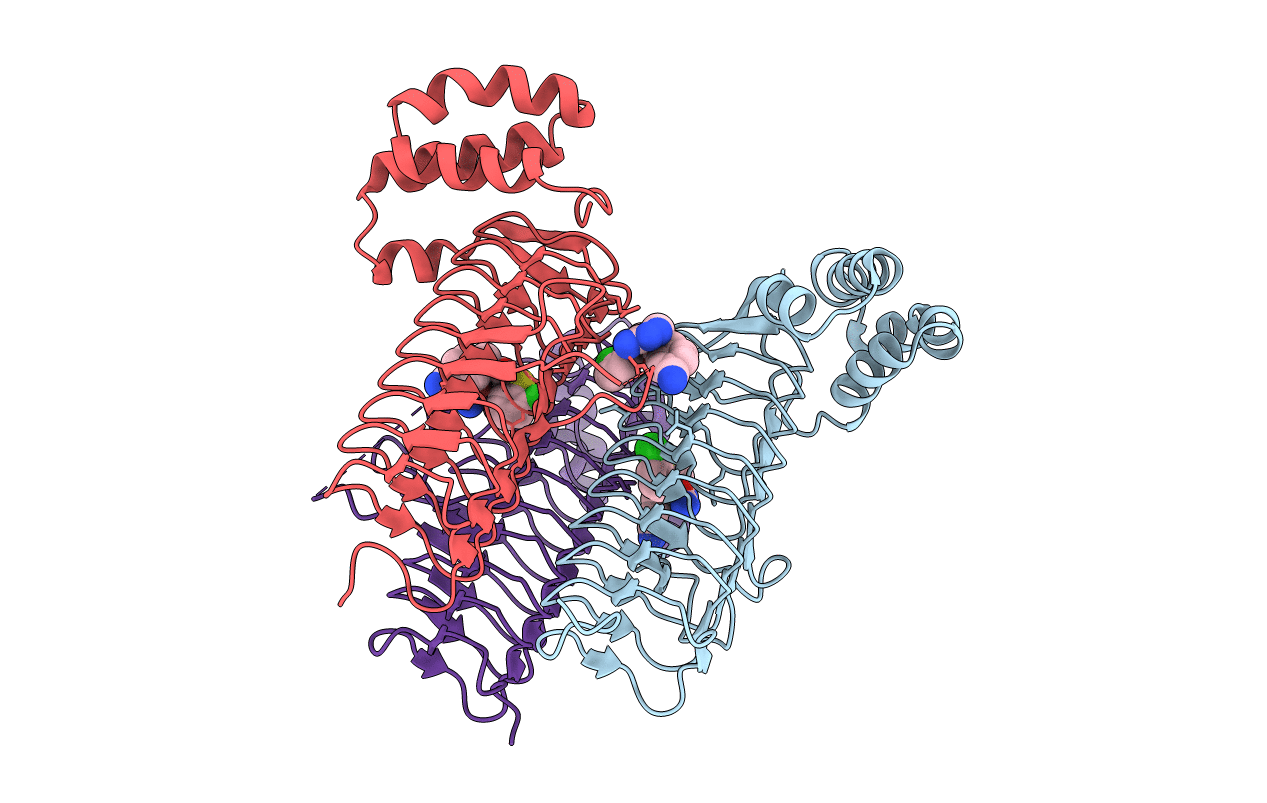
Crystal Structure Of Pseudomonas Aeruginosa Lpxa In Complex With Compound 93
Organism: Pseudomonas aeruginosa (strain pa7)
Method: X-RAY DIFFRACTION Resolution:1.72 Å Release Date: 2021-10-13 Classification: TRANSFERASE Ligands: VFW |
 |
Crystal Structure Of Pseudomonas Aeruginosa Lpxa In Complex With Compound 3
Organism: Pseudomonas aeruginosa (strain pa7)
Method: X-RAY DIFFRACTION Resolution:1.92 Å Release Date: 2021-10-13 Classification: TRANSFERASE Ligands: VFN, CL |
 |
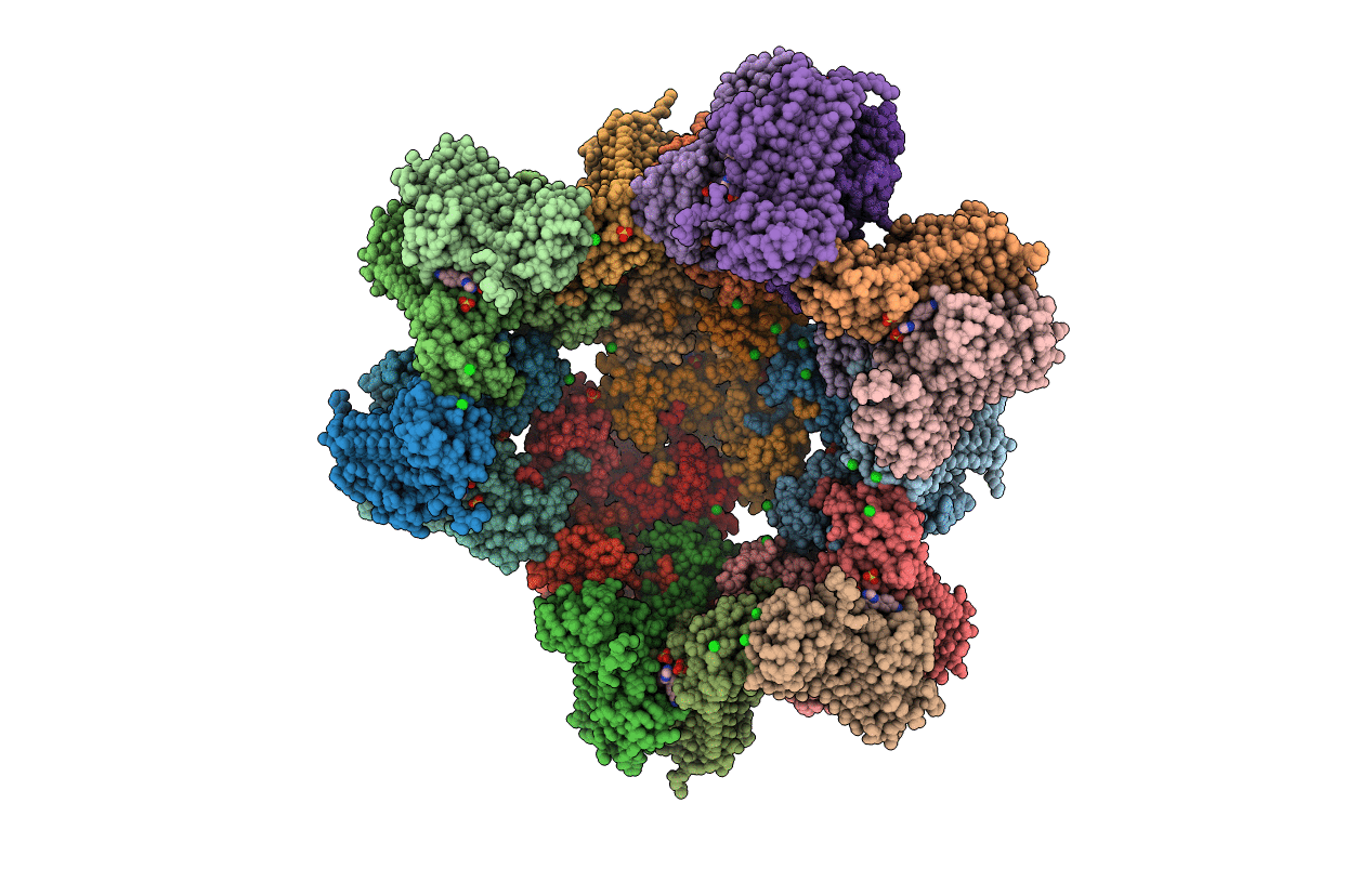
Crystal Structure Of Pseudomonas Aeruginosa Lpxa In Complex With Compound 3
Organism: Pseudomonas aeruginosa (strain pa7)
Method: X-RAY DIFFRACTION Resolution:2.89 Å Release Date: 2021-10-13 Classification: TRANSFERASE Ligands: VFN, SO4 |
 |
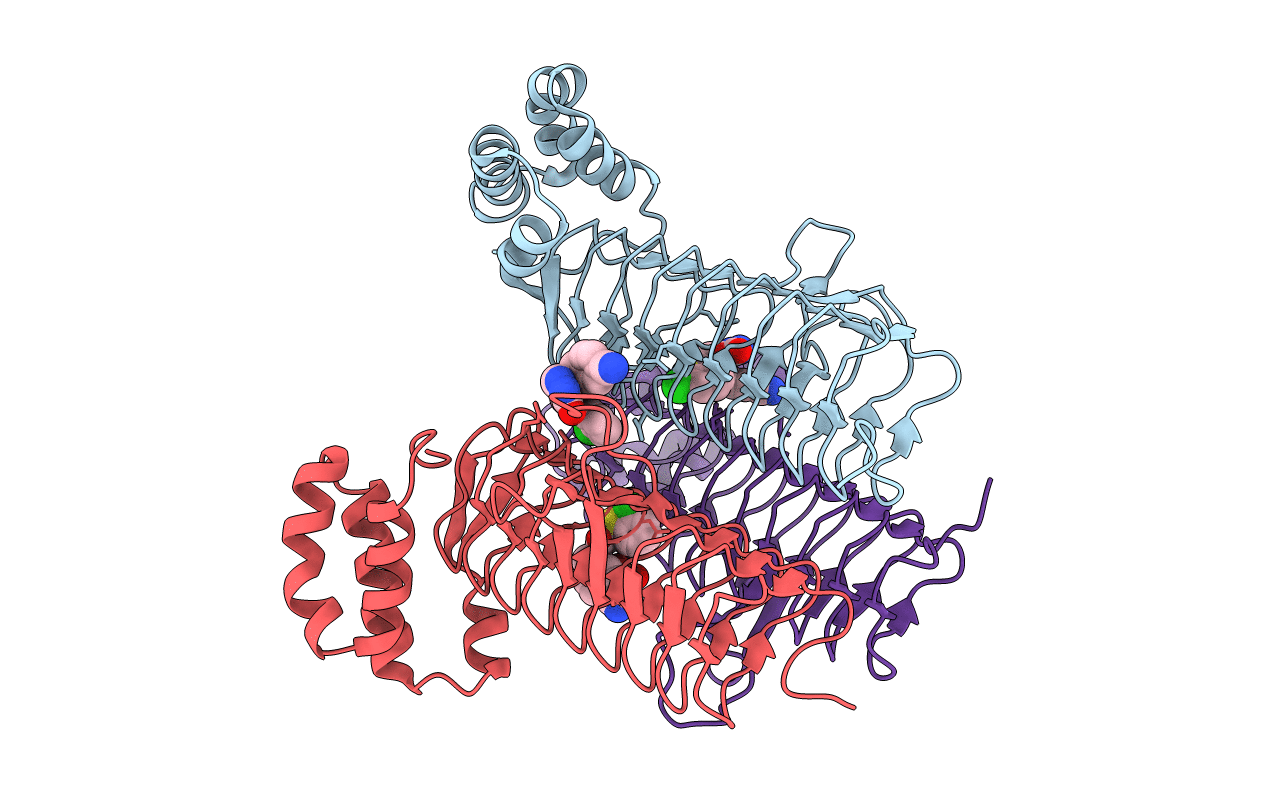
Crystal Structure Of Pseudomonas Aeruginosa Lpxa In Complex With Compound 14
Organism: Pseudomonas aeruginosa (strain pa7)
Method: X-RAY DIFFRACTION Resolution:2.74 Å Release Date: 2021-10-13 Classification: TRANSFERASE Ligands: VFT, CL, SO4 |
 |
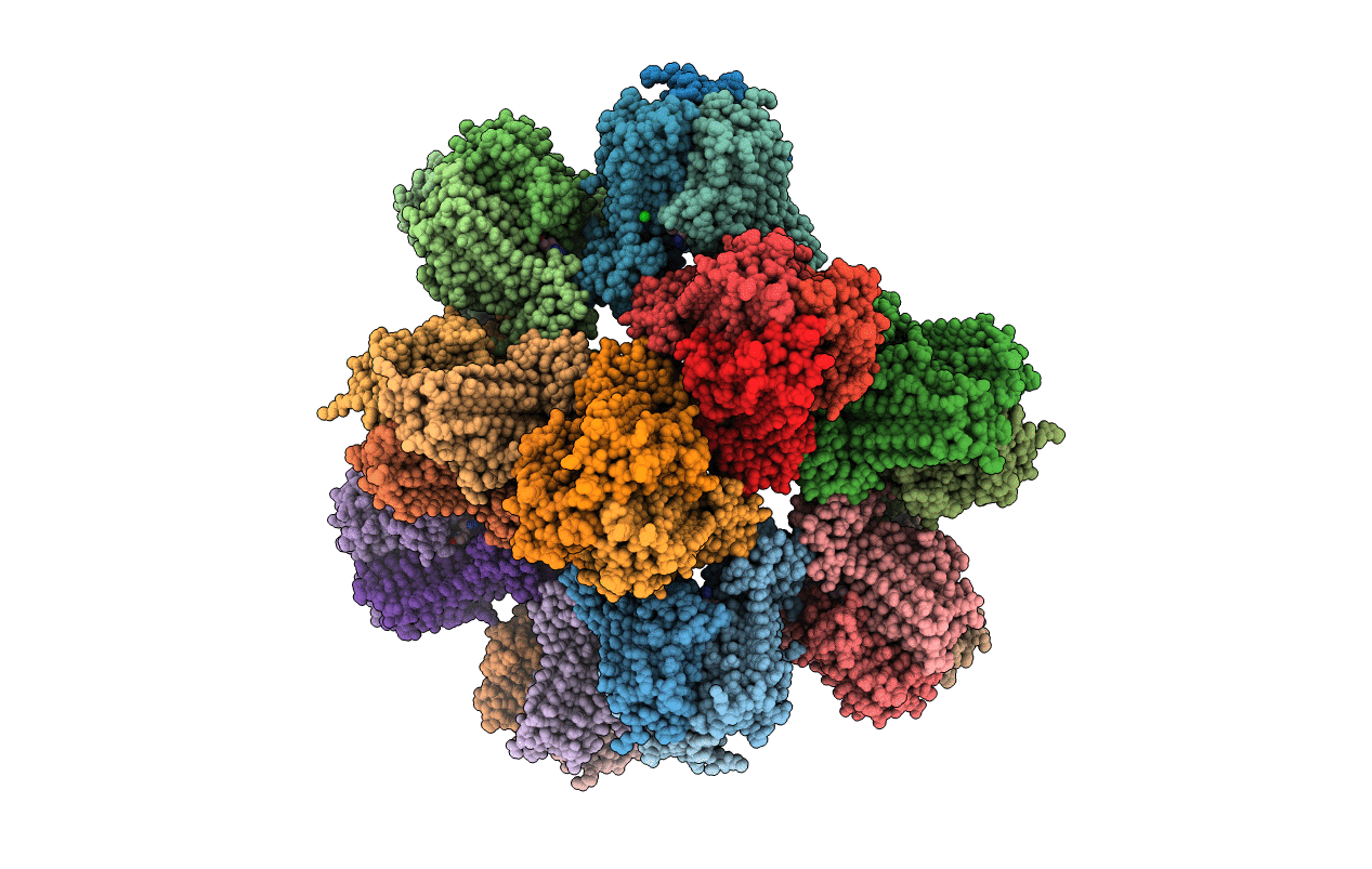
Crystal Structure Of Pseudomonas Aeruginosa Lpxa In Complex With Compound 6
Organism: Pseudomonas aeruginosa (strain pa7)
Method: X-RAY DIFFRACTION Resolution:2.00 Å Release Date: 2021-10-06 Classification: TRANSFERASE Ligands: VGQ, CL |
 |
Crystal Structure Of Pseudomonas Aeruginosa Lpxa In Complex With Compound 45
Organism: Pseudomonas aeruginosa
Method: X-RAY DIFFRACTION Resolution:3.58 Å Release Date: 2021-10-06 Classification: TRANSFERASE Ligands: CL, SO4, VFZ |
 |
Organism: Escherichia coli
Method: X-RAY DIFFRACTION Resolution:1.84 Å Release Date: 2021-10-06 Classification: TRANSFERASE Ligands: VFE, NA |
 |
Organism: Pseudomonas aeruginosa
Method: X-RAY DIFFRACTION Resolution:1.90 Å Release Date: 2017-03-29 Classification: HYDROLASE Ligands: ZN, 8Q8, CL |
 |
Crystal Structure Of The Atp Binding Domain Of S. Aureus Gyrb Complexed With A Fragment
Organism: Staphylococcus aureus
Method: X-RAY DIFFRACTION Resolution:1.20 Å Release Date: 2016-02-03 Classification: ISOMERASE/ISOMERASE INHIBITOR Ligands: EVO, SO4, MPD |
 |
Crystal Structure Of The Atp Binding Domain Of S. Aureus Gyrb Complexed With A Fragment
Organism: Staphylococcus aureus
Method: X-RAY DIFFRACTION Resolution:1.45 Å Release Date: 2016-02-03 Classification: ISOMERASE Ligands: MPD, CL, 54X, MG |
 |
Crystal Structure Of The Atp Binding Domain Of S. Aureus Gyrb Complexed With A Fragment
Organism: Staphylococcus aureus
Method: X-RAY DIFFRACTION Resolution:1.48 Å Release Date: 2016-02-03 Classification: ISOMERASE/ISOMERASE INHIBITOR Ligands: 55D, MG, CL, MPD |
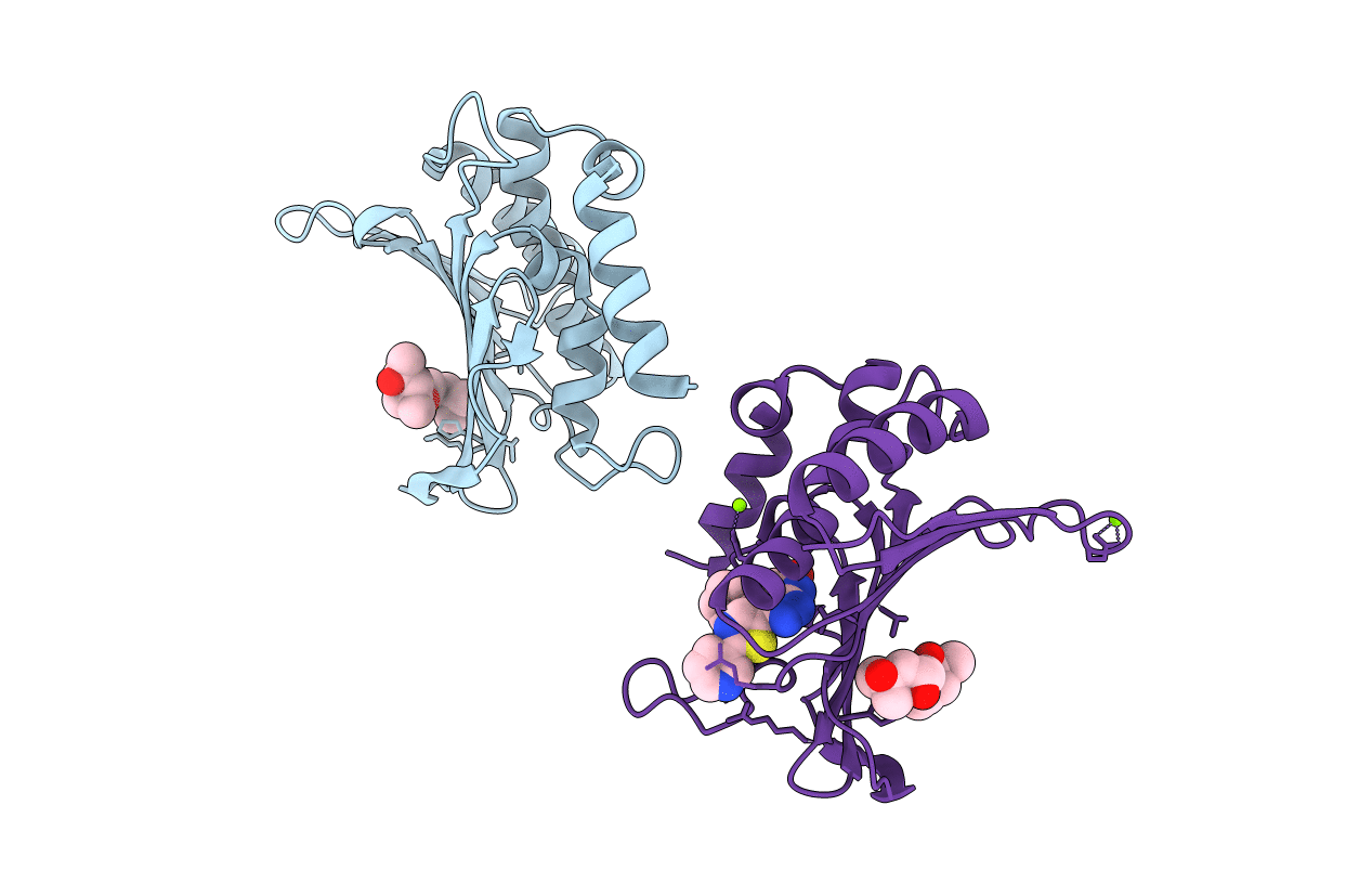 |
Crystal Structure Of The Atp Binding Domain Of S. Aureus Gyrb Complexed With A Fragment
Organism: Staphylococcus aureus
Method: X-RAY DIFFRACTION Resolution:1.60 Å Release Date: 2016-02-03 Classification: ISOMERASE/ISOMERASE INHIBITOR Ligands: MPD, 55G, MG |
 |
Crystal Structure Of The Atp Binding Domain Of S. Aureus Gyrb Complexed With A Fragment
Organism: Staphylococcus aureus
Method: X-RAY DIFFRACTION Resolution:1.60 Å Release Date: 2016-02-03 Classification: ISOMERASE/ISOMERASE INHIBITOR Ligands: MPD, CL, MG, 55H |
 |
Organism: Homo sapiens
Method: X-RAY DIFFRACTION Resolution:2.22 Å Release Date: 2011-06-08 Classification: HYDROLASE Ligands: GOL, DW0 |
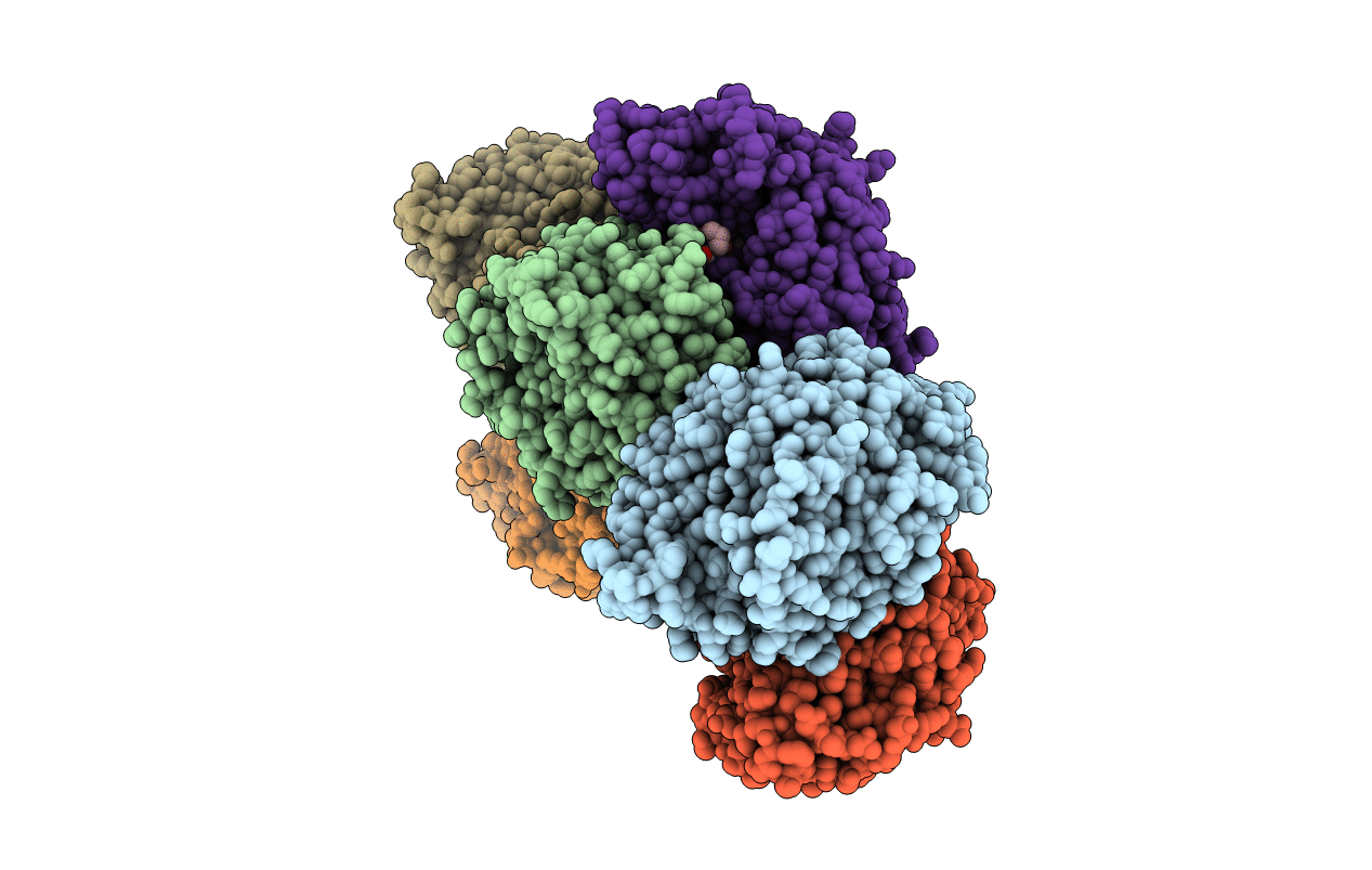 |
Organism: Homo sapiens
Method: X-RAY DIFFRACTION Resolution:2.25 Å Release Date: 2011-06-08 Classification: HYDROLASE Ligands: GOL, CX9 |
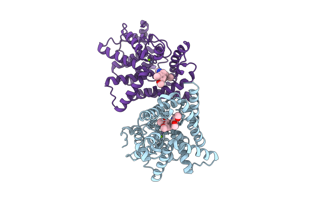 |
Human Pde-Papaverine Complex Obtained By Ligand Soaking Of Cross- Linked Protein Crystals
Organism: Homo sapiens
Method: X-RAY DIFFRACTION Resolution:2.80 Å Release Date: 2009-07-28 Classification: HYDROLASE Ligands: EV1, ZN, MG |
 |
Crystal Structure Of Aspergillus Fumigatus Chitinase B1 In Complex With Dimethylguanylurea
Organism: Aspergillus fumigatus
Method: X-RAY DIFFRACTION Resolution:2.20 Å Release Date: 2008-03-25 Classification: HYDROLASE/HYDROLASE inhibitor Ligands: SO4, XRG |

