
Search Count: 52
 |
Organism: Homo sapiens
Method: ELECTRON MICROSCOPY Release Date: 2025-10-08 Classification: STRUCTURAL PROTEIN Ligands: ADP, MG, PO4 |
 |
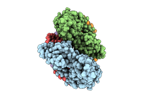
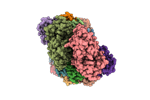
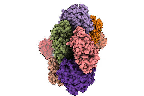
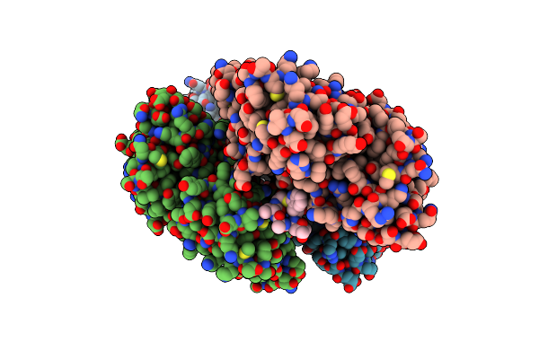
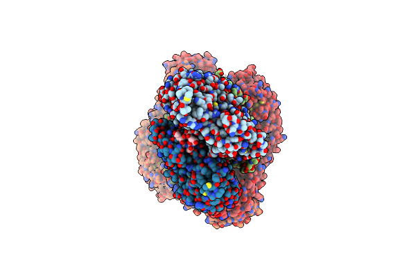
Cryo-Em Structure Of The Actin Filament Bound By A Single Coronin-1B Molecule.
Organism: Homo sapiens
Method: ELECTRON MICROSCOPY Release Date: 2025-10-08 Classification: STRUCTURAL PROTEIN Ligands: ADP, MG, PO4 |
 |
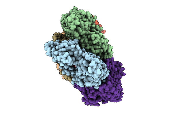
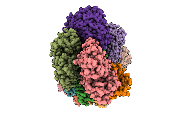
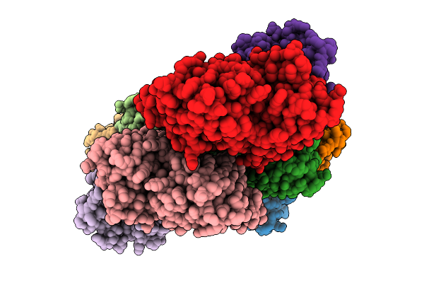
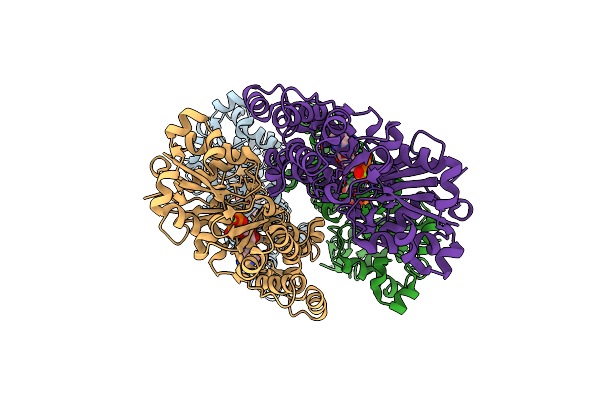
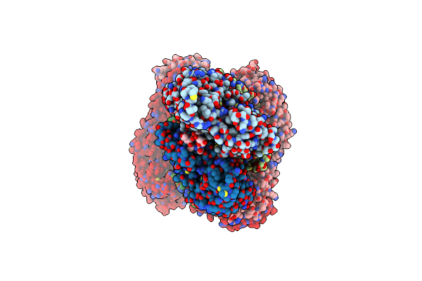
Cryo-Em Structure Of The Fully Coronin-1B-Decorated Actin Filament In The Adp State.
Organism: Homo sapiens
Method: ELECTRON MICROSCOPY Release Date: 2025-10-08 Classification: STRUCTURAL PROTEIN Ligands: ADP, MG |
 |
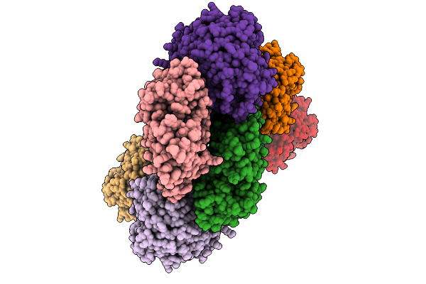
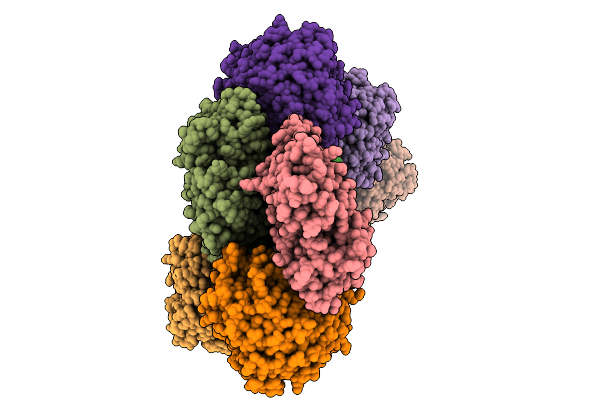
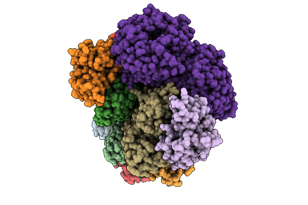
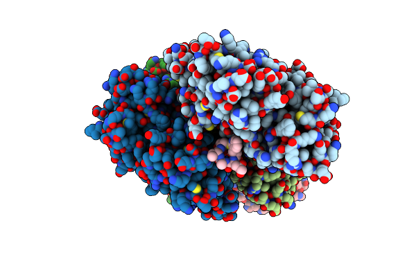
Cryo-Em Structure Of The Fully Coronin-1B-Decorated Actin Filament In The Adp State.
Organism: Homo sapiens
Method: ELECTRON MICROSCOPY Release Date: 2025-10-08 Classification: STRUCTURAL PROTEIN Ligands: ADP, MG |
 |
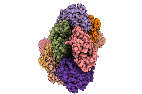
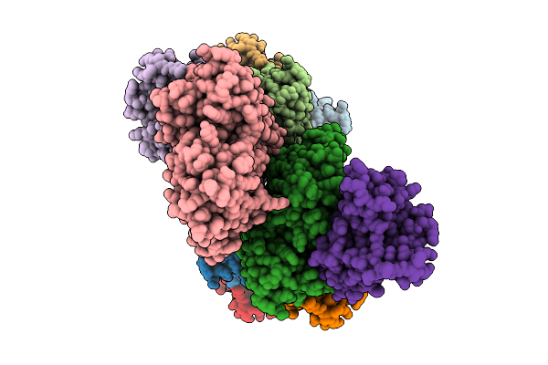
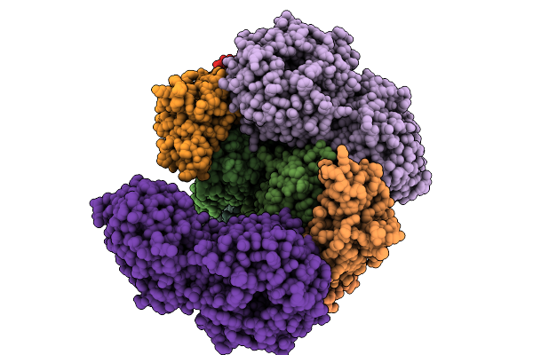
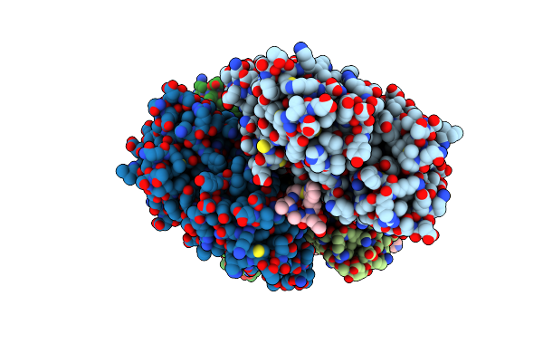
Cryo-Em Structure Of The Fully Cofilin-1-Decorated Actin Filament (Cofilactin)
Organism: Homo sapiens
Method: ELECTRON MICROSCOPY Release Date: 2025-10-08 Classification: STRUCTURAL PROTEIN Ligands: ADP, MG |
 |
Cryo-Em Structure Of The Actin Filament Hetero-Decorated By Coronin-1 And Cofilin-1 On Separate Actin Strands
Organism: Homo sapiens
Method: ELECTRON MICROSCOPY Release Date: 2025-10-08 Classification: STRUCTURAL PROTEIN Ligands: ADP, MG |
 |
Cryo-Em Structure Of The Actin Filament Hetero-Decorated By Coronin-1 And Cofilin-1, Strand Boundary
Organism: Homo sapiens
Method: ELECTRON MICROSCOPY Release Date: 2025-10-08 Classification: STRUCTURAL PROTEIN Ligands: ADP, MG |
 |
Cryo-Em Structure Of The Cofilactin Filament Core At 2.3 Angstrom Resolution.
Organism: Homo sapiens
Method: ELECTRON MICROSCOPY Release Date: 2025-10-08 Classification: STRUCTURAL PROTEIN Ligands: ADP, MG |
 |
Cryo-Em Structure Of The Coronin-1B-Decorated Actin Filament Bound By One Cofilin-1 Molecule (Crosslinked)
Organism: Homo sapiens
Method: ELECTRON MICROSCOPY Release Date: 2025-10-08 Classification: STRUCTURAL PROTEIN Ligands: ADP, MG |
 |
Organism: Homo sapiens
Method: ELECTRON MICROSCOPY Release Date: 2025-10-08 Classification: STRUCTURAL PROTEIN Ligands: ADP, MG |
 |
Organism: Homo sapiens
Method: ELECTRON MICROSCOPY Release Date: 2025-10-08 Classification: STRUCTURAL PROTEIN Ligands: MG, ADP |
 |
Organism: Homo sapiens
Method: ELECTRON MICROSCOPY Release Date: 2025-10-08 Classification: STRUCTURAL PROTEIN Ligands: MG, ADP |
 |
Cryo-Em Structure Of The Phalloidin-Bound Pointed End Of The Actin Filament.
Organism: Homo sapiens, Amanita phalloides
Method: ELECTRON MICROSCOPY Release Date: 2024-09-11 Classification: STRUCTURAL PROTEIN Ligands: ADP, MG, PO4 |
 |
Organism: Oryctolagus cuniculus
Method: ELECTRON MICROSCOPY Release Date: 2024-09-11 Classification: STRUCTURAL PROTEIN Ligands: ADP, MG |
 |
Structure Of The Dnase I- And Phalloidin-Bound Pointed End Of F-Actin (Conformer 1)
Organism: Bos taurus, Amanita phalloides
Method: ELECTRON MICROSCOPY Release Date: 2024-09-11 Classification: STRUCTURAL PROTEIN Ligands: ADP, MG, PO4 |
 |
Structure Of The Dnase I- And Phalloidin-Bound Pointed End Of F-Actin (Conformer 2).
Organism: Bos taurus, Amanita phalloides
Method: ELECTRON MICROSCOPY Release Date: 2024-09-11 Classification: STRUCTURAL PROTEIN Ligands: ADP, MG, PO4 |
 |
Crystal Structure Of Human Casein Kinase Ii Subunit Alpha (Ck2A1) In Complex With Tr06772818
Organism: Homo sapiens
Method: X-RAY DIFFRACTION Release Date: 2024-09-11 Classification: TRANSFERASE Ligands: SO4, A1IG6 |
 |
Crystal Structure Of Human Casein Kinase Ii Subunit Alpha (Ck2A1) In Complex With 5-((4-((2-Aminoethyl)(Ethyl)Amino)-3-(4H-1,2,4-Triazol-4-Yl)Phenyl)Amino)-7-(Cyclopropylamino)Pyrazolo[1,5-A]Pyrimidine-3-Carbonitrile
Organism: Homo sapiens
Method: X-RAY DIFFRACTION Resolution:2.18 Å Release Date: 2024-07-31 Classification: TRANSFERASE Ligands: SO4, A1H8C, EDO |
 |
Structure Of The Formin Cdc12 Bound To The Barbed End Of Phalloidin-Stabilized F-Actin.
Organism: Homo sapiens, Schizosaccharomyces pombe, Amanita phalloides
Method: ELECTRON MICROSCOPY Release Date: 2024-04-10 Classification: STRUCTURAL PROTEIN Ligands: ADP, MG, PO4 |
 |
Structure Of The F-Actin Barbed End Bound By Cdc12 And Profilin (Ring Complex) At A Resolution Of 6.3 Angstrom
Organism: Homo sapiens, Schizosaccharomyces pombe, Amanita phalloides
Method: ELECTRON MICROSCOPY Release Date: 2024-04-10 Classification: STRUCTURAL PROTEIN Ligands: ADP, MG, PO4 |