
Search Count: 5,384
 |
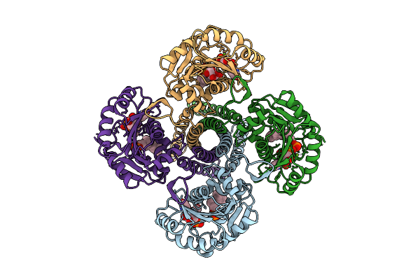
Organism: African swine fever virus
Method: ELECTRON MICROSCOPY Release Date: 2025-12-31 Classification: DNA BINDING PROTEIN |
 |
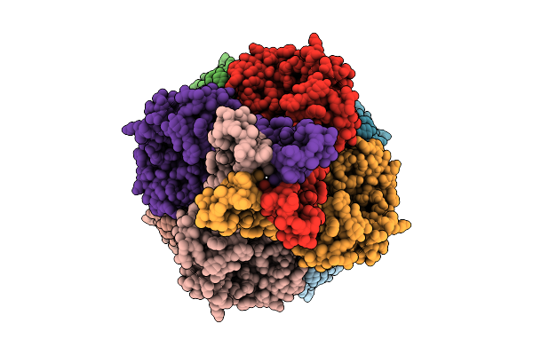
Organism: Asfarviridae
Method: ELECTRON MICROSCOPY Release Date: 2025-12-31 Classification: DNA BINDING PROTEIN/DNA Ligands: DCP, MG |
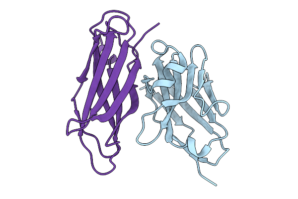 |
Crystal Structure Of Malaria Transmission-Blocking Antigen Pfhap2 Domain 3 In Complex With Nanobody Wnb 334
Organism: Vicugna pacos, Plasmodium falciparum
Method: X-RAY DIFFRACTION Release Date: 2025-12-31 Classification: PROTEIN BINDING |
 |
Crystal Structure Of An Allosteric Inhibitor Bound To Human Ripk1 Kinase Domain
Organism: Homo sapiens
Method: X-RAY DIFFRACTION Release Date: 2025-12-24 Classification: IMMUNE SYSTEM Ligands: A1IX7, IOD, 1HX |
 |
Crystal Structure Of An Allosteric Inhibitor Bound To Human Ripk1 Kinase Domain
Organism: Homo sapiens
Method: X-RAY DIFFRACTION Release Date: 2025-12-24 Classification: IMMUNE SYSTEM Ligands: A1IX9, IOD, PEG |
 |
Cryo-Em Structure Of The Glycosyltransferase Gtrb In The Substrate-Bound State
Organism: Synechocystis sp. pcc 6803 substr. kazusa
Method: ELECTRON MICROSCOPY Release Date: 2025-12-17 Classification: MEMBRANE PROTEIN Ligands: UPG, 5TR, MG |
 |
Cryo-Em Structure Of The Glycosyltransferase Gtrb In The Pre-Catalysis And Product-Bound State
Organism: Synechocystis sp. pcc 6803 substr. kazusa
Method: ELECTRON MICROSCOPY Release Date: 2025-12-17 Classification: MEMBRANE PROTEIN Ligands: UPG, 5TR, MG, UDP, A1B94 |
 |
Cryo-Em Structure Of The Glycosyltransferase Gtrb In The Apo State (Octamer Volume)
Organism: Synechocystis sp. pcc 6803 substr. kazusa
Method: ELECTRON MICROSCOPY Release Date: 2025-12-17 Classification: MEMBRANE PROTEIN |
 |
Organism: Synechocystis sp. pcc 6803 substr. kazusa
Method: ELECTRON MICROSCOPY Release Date: 2025-12-17 Classification: MEMBRANE PROTEIN Ligands: UPG, 5TR, MG |
 |
Cryo-Em Structure Of The Glycosyltransferase Gtrb In The Pre-Intermediate State
Organism: Synechocystis sp. pcc 6803 substr. kazusa
Method: ELECTRON MICROSCOPY Release Date: 2025-12-17 Classification: MEMBRANE PROTEIN Ligands: UPG, 5TR, MG |
 |
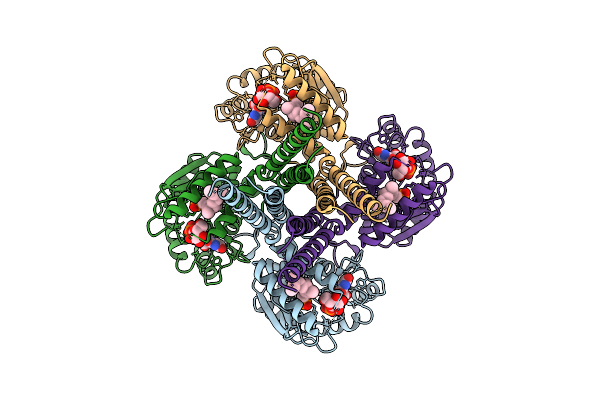
Organism: Gut metagenome
Method: ELECTRON MICROSCOPY Release Date: 2025-12-10 Classification: CYTOSOLIC PROTEIN |
 |
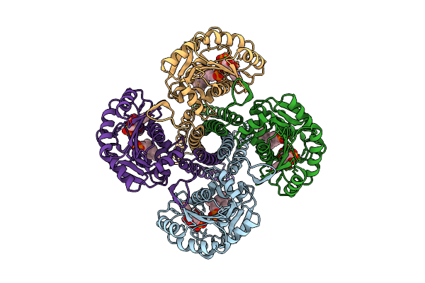
Organism: Gut metagenome
Method: ELECTRON MICROSCOPY Release Date: 2025-12-10 Classification: HYDROLASE Ligands: PO4, BGC |
 |
Organism: Homo sapiens
Method: X-RAY DIFFRACTION Release Date: 2025-12-03 Classification: UNKNOWN FUNCTION |
 |
Organism: Homo sapiens
Method: X-RAY DIFFRACTION Release Date: 2025-12-03 Classification: UNKNOWN FUNCTION |
 |
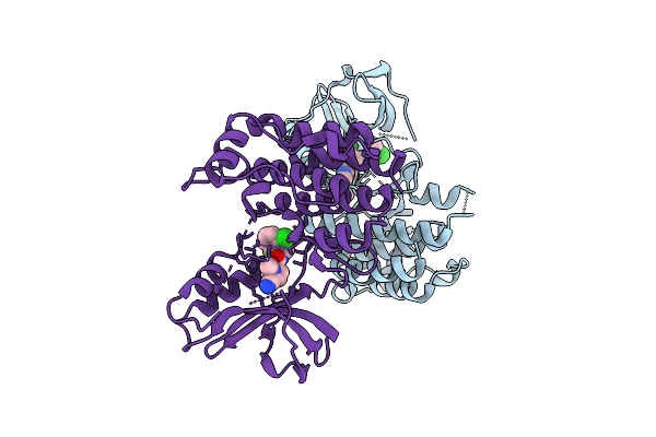
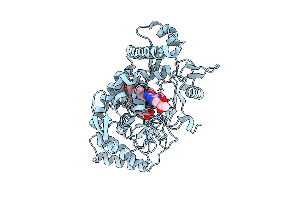
Structure Of Human Neuronal Nitric Oxide Synthase R354A/G357D Mutant Heme Domain Bound With 7-(3-Aminomethyl)Phenyl-6-Fluoro-4-Methylquinoin-2-Amine
Organism: Homo sapiens
Method: X-RAY DIFFRACTION Release Date: 2025-12-03 Classification: OXIDOREDUCTASE Ligands: HEM, H4B, A1BUD, GOL, ZN |
 |
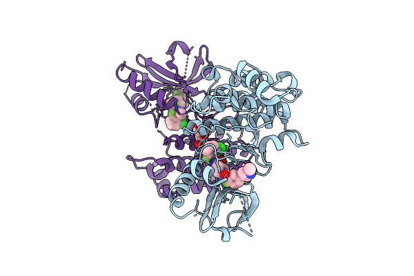
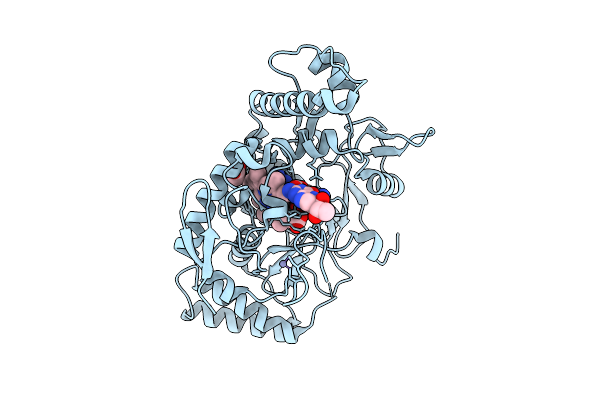
Structure Of Human Neuronal Nitric Oxide Synthase R354A/G357D Mutant Heme Domain Bound With 2-(2-Amino-6-Fluoro-4-Methylquinolin-7-Yl)-5-(Aminomethyl)Phenol
Organism: Homo sapiens
Method: X-RAY DIFFRACTION Release Date: 2025-12-03 Classification: OXIDOREDUCTASE Ligands: HEM, H4B, A1BUF, GOL, ZN |
 |
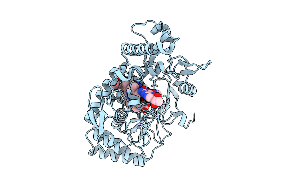
Structure Of Human Neuronal Nitric Oxide Synthase R354A/G357D Mutant Heme Domain Bound 2-(2-Amino-6-Fluoro-4-Methylquinolin-7-Yl)-5-(2-Aminoethyl)Phenol
Organism: Homo sapiens
Method: X-RAY DIFFRACTION Release Date: 2025-12-03 Classification: OXIDOREDUCTASE Ligands: HEM, H4B, A1BUH, GOL, ZN |
 |
Structure Of Rat Neuronal Nitric Oxide Synthase R349A Mutant Heme Domain Bound With 7-(3-Aminomethyl)Phenyl-6-Fluoro-4-Methylquinoin-2-Amine
Organism: Rattus norvegicus
Method: X-RAY DIFFRACTION Release Date: 2025-12-03 Classification: OXIDOREDUCTASE Ligands: HEM, H4B, A1BUD, ACT, ZN |
 |
Structure Of Rat Neuronal Nitric Oxide Synthase R349A Mutant Heme Domain Bound With 2-(2-Amino-6-Fluoro-4-Methylquinolin-7-Yl)-5-(Aminomethyl)Phenol
Organism: Rattus norvegicus
Method: X-RAY DIFFRACTION Release Date: 2025-12-03 Classification: OXIDOREDUCTASE Ligands: HEM, H4B, ACT, ZN, A1BUF |
 |
Structure Of Rat Neuronal Nitric Oxide Synthase R349A Mutant Heme Domain Bound 2-(2-Amino-6-Fluoro-4-Methylquinolin-7-Yl)-5-(2-Aminoethyl)Phenol
Organism: Rattus norvegicus
Method: X-RAY DIFFRACTION Release Date: 2025-12-03 Classification: OXIDOREDUCTASE Ligands: HEM, H4B, ACT, ZN, A1BUH |