
Search Count: 33
 |
Organism: Salmonella enterica subsp. enterica serovar typhimurium str. 14028s
Method: X-RAY DIFFRACTION Release Date: 2025-10-22 Classification: PHOTOSYNTHESIS Ligands: A1EKZ, SO4 |
 |
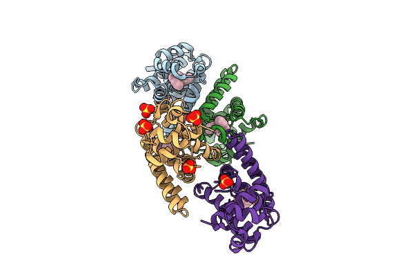
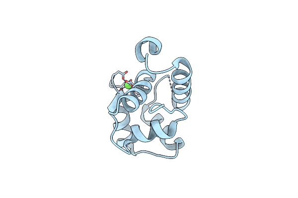
Crystal Structure Of F10 Core Protein From Monkeypox Virus Reveals Its Potential Inhibitors
Organism: Monkeypox virus
Method: X-RAY DIFFRACTION Resolution:1.51 Å Release Date: 2024-11-13 Classification: VIRAL PROTEIN |
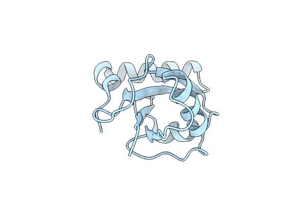 |
Organism: Pyricularia oryzae
Method: X-RAY DIFFRACTION Resolution:2.50 Å Release Date: 2024-09-04 Classification: APOPTOSIS |
 |
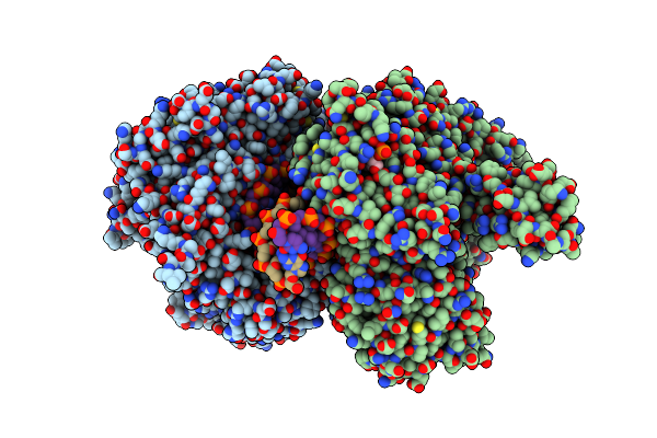
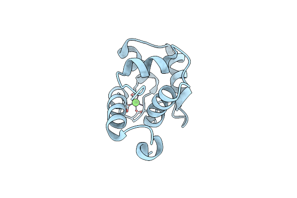
Organism: Homo sapiens
Method: X-RAY DIFFRACTION Resolution:1.74 Å Release Date: 2024-05-01 Classification: HYDROLASE/ONCOPROTEIN Ligands: GTP, MG, UNX |
 |
Organism: Rubella virus (strain ra27/3 vaccine)
Method: X-RAY DIFFRACTION Resolution:1.64 Å Release Date: 2022-07-13 Classification: HYDROLASE Ligands: GOL, EDO, PEG, ZN |
 |
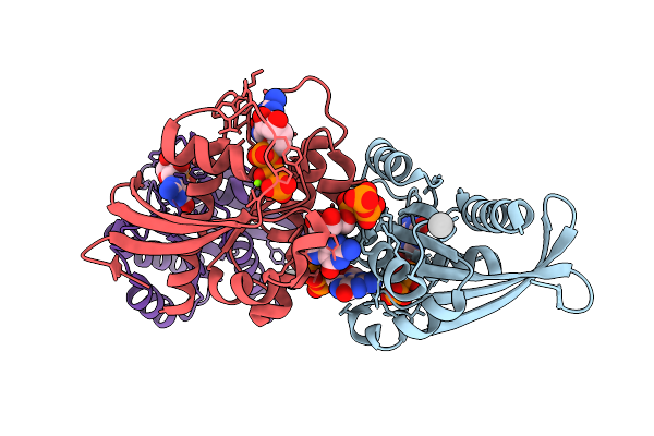
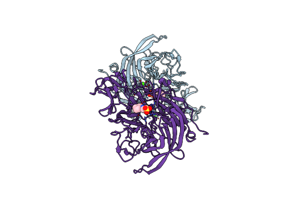
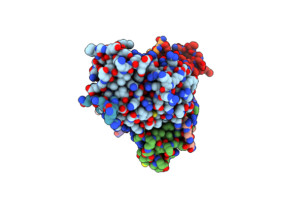
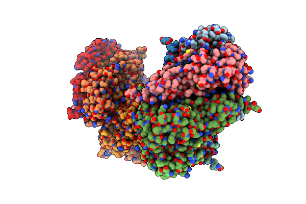
Crystal Structure Of Replisomal Dimer Of Dna Polymerase From Bacteriophage Rb69 With Dna Duplexes
Organism: Enterobacteria phage rb69, Synthetic construct
Method: X-RAY DIFFRACTION Resolution:2.20 Å Release Date: 2021-07-14 Classification: TRANSFERASE/DNA Ligands: DUP, CA, 5GP, MG |
 |
Organism: Streptococcus mutans
Method: X-RAY DIFFRACTION Resolution:1.50 Å Release Date: 2019-02-06 Classification: SIGNALING PROTEIN |
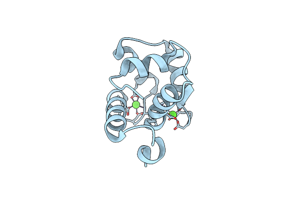 |
Organism: Homo sapiens
Method: X-RAY DIFFRACTION Resolution:1.86 Å Release Date: 2017-09-13 Classification: METAL BINDING PROTEIN Ligands: CA |
 |
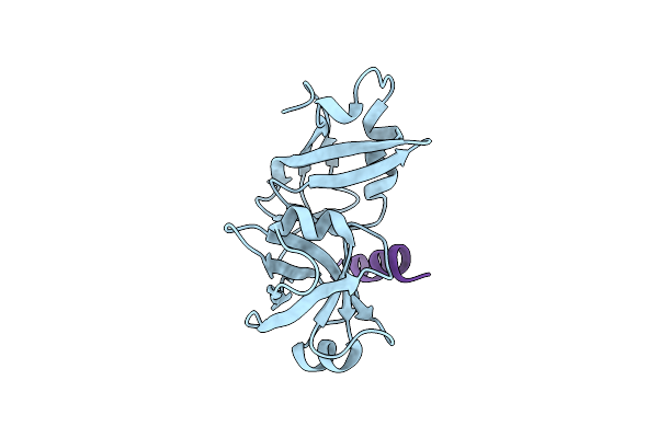
Crystal Structure Of The Catalytic Domain Of Clostridium Perfringens Neuraminidase (Nani) In Complex With A Ches
Organism: Clostridium perfringens atcc 13124
Method: X-RAY DIFFRACTION Resolution:1.24 Å Release Date: 2017-03-29 Classification: HYDROLASE Ligands: NHE, CA |
 |
Organism: Homo sapiens
Method: X-RAY DIFFRACTION Resolution:1.85 Å Release Date: 2016-12-28 Classification: METAL BINDING PROTEIN Ligands: CA |
 |
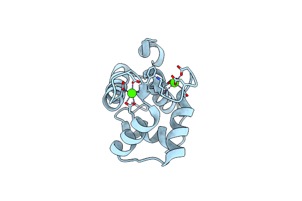
Organism: Homo sapiens
Method: X-RAY DIFFRACTION Resolution:1.95 Å Release Date: 2016-12-28 Classification: METAL BINDING PROTEIN Ligands: CA |
 |
Organism: Homo sapiens
Method: X-RAY DIFFRACTION Resolution:1.94 Å Release Date: 2016-12-28 Classification: METAL BINDING PROTEIN Ligands: CA |
 |
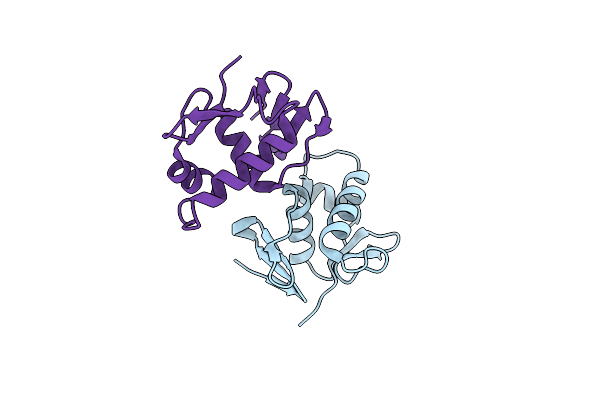
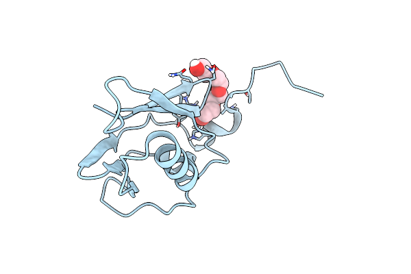
Structural Insights Into The Interaction Of P97 N-Terminal Domain And Shp Motif In Derlin-1 Rhomboid Pseudoprotease
Organism: Homo sapiens
Method: X-RAY DIFFRACTION Resolution:2.25 Å Release Date: 2016-11-09 Classification: HYDROLASE/TRANSPORT PROTEIN |
 |
Structural Insights Into The Interaction Of P97 N-Terminus Domain And Vbm Motif In Rhomboid Protease, Rhbdl4
Organism: Homo sapiens
Method: X-RAY DIFFRACTION Resolution:1.88 Å Release Date: 2016-09-14 Classification: HYDROLASE |
 |
Organism: Homo sapiens
Method: X-RAY DIFFRACTION Resolution:2.80 Å Release Date: 2016-02-03 Classification: TRANSFERASE |
 |
Crystal Structure Of Human Quinolinate Phosphoribosyltransferase In Complex With The Reactant Quinolinate
Organism: Homo sapiens
Method: X-RAY DIFFRACTION Resolution:3.09 Å Release Date: 2016-02-03 Classification: TRANSFERASE Ligands: NTM |
 |
Crystal Structure Of Human Quinolinate Phosphoribosyltransferase In Complex With The Product Nicotinate Mononucleotide
Organism: Homo sapiens
Method: X-RAY DIFFRACTION Resolution:2.60 Å Release Date: 2016-02-03 Classification: TRANSFERASE Ligands: NCN |
 |
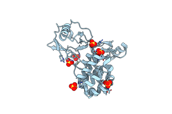
Crystal Structure Of The N-Terminal Domain Of The Human Mitochondrial Calcium Uniporter Fused With T4 Lysozyme
Organism: Enterobacteria phage t4, Homo sapiens
Method: X-RAY DIFFRACTION Resolution:1.80 Å Release Date: 2015-09-16 Classification: TRANSPORT PROTEIN Ligands: SO4 |
 |
Crystal Structure Of The N-Terminal Domain Of The Human Mitochondrial Calcium Uniporter
Organism: Homo sapiens
Method: X-RAY DIFFRACTION Resolution:1.50 Å Release Date: 2015-09-16 Classification: TRANSPORT PROTEIN Ligands: PG4 |
 |
Crystal Structure Of The N-Terminal Domain Single Mutant (S92A) Of The Human Mitochondrial Calcium Uniporter Fused With T4 Lysozyme
Organism: Enterobacteria phage t4, Homo sapiens
Method: X-RAY DIFFRACTION Resolution:2.75 Å Release Date: 2015-09-16 Classification: TRANSPORT PROTEIN Ligands: SO4 |