
Search Count: 2,613
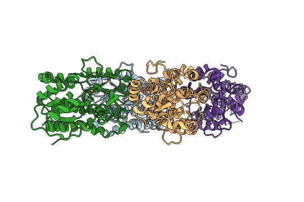 |
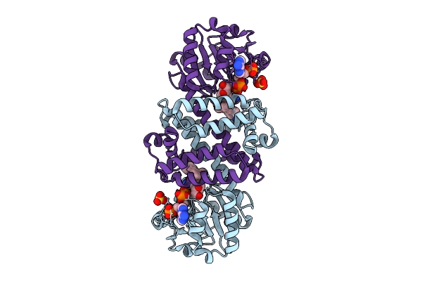
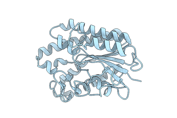
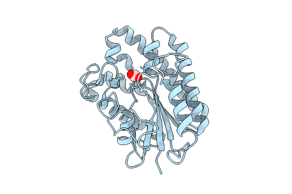
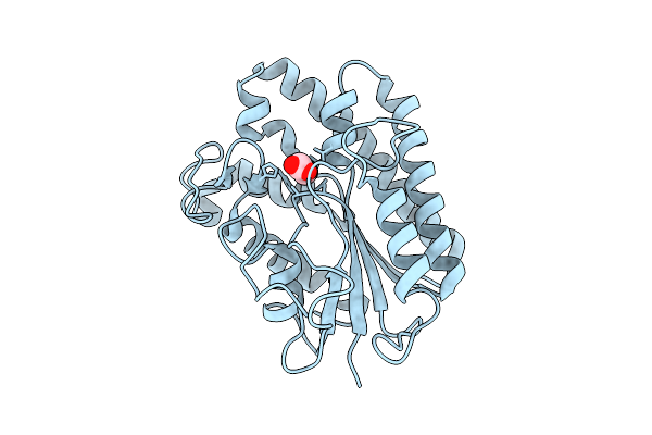
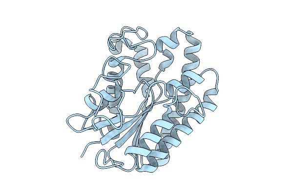
Organism: Yersinia ruckeri
Method: X-RAY DIFFRACTION Release Date: 2025-11-05 Classification: METAL BINDING PROTEIN Ligands: FE |
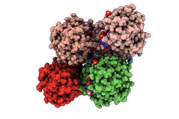 |
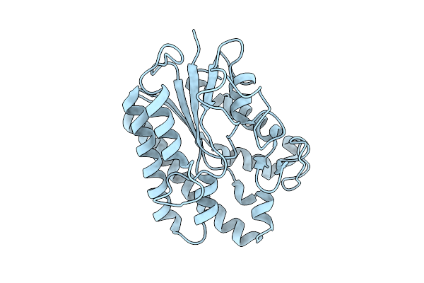
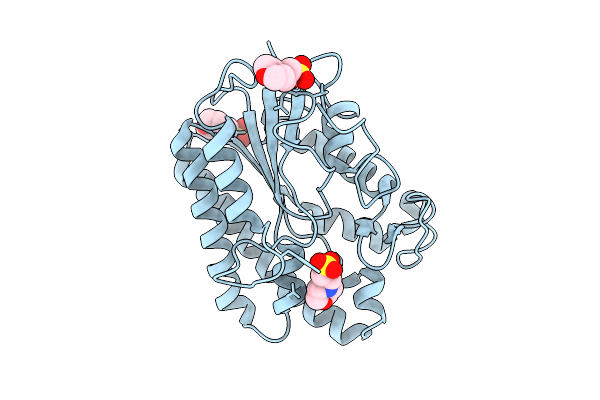
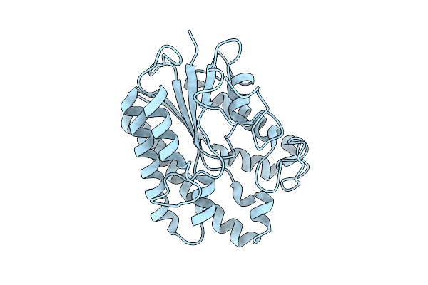
Organism: Yersinia ruckeri
Method: X-RAY DIFFRACTION Release Date: 2025-11-05 Classification: METAL BINDING PROTEIN Ligands: FE, A1I15, SO4 |
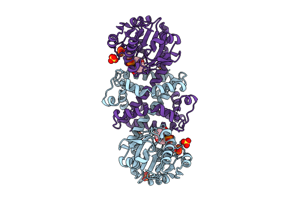 |
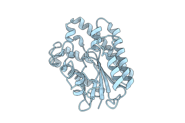
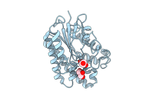
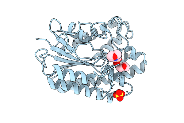
Organism: Kribbella flavida
Method: X-RAY DIFFRACTION Release Date: 2025-11-05 Classification: OXIDOREDUCTASE Ligands: NAP, SO4 |
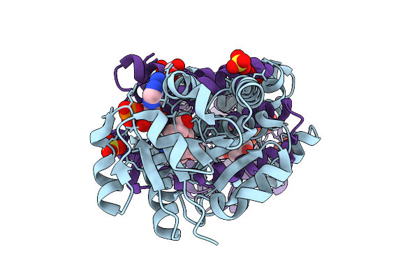 |
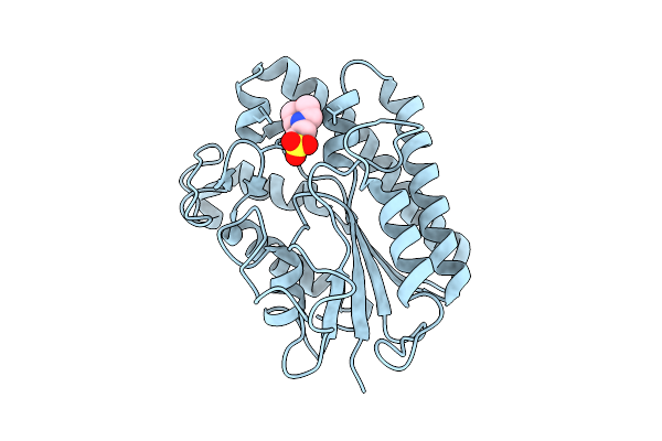
Imine Reductase Ir91 From Kribbella Flavida With Nadp+ And 5-Methoxy-2-Tetralone
Organism: Kribbella flavida
Method: X-RAY DIFFRACTION Release Date: 2025-11-05 Classification: OXIDOREDUCTASE Ligands: SO4, NAP, A1JDH, EDO |
 |
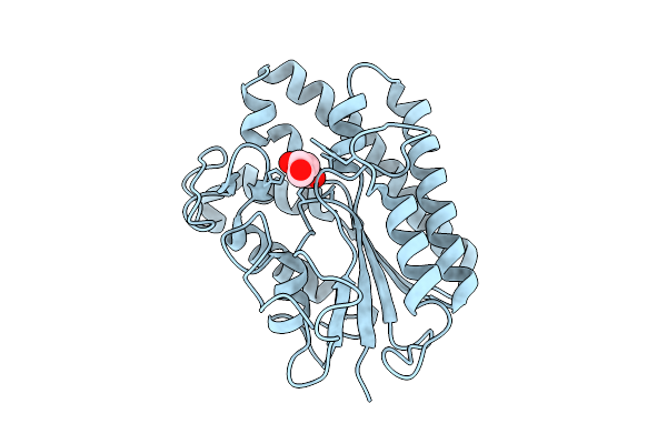
Imine Reductase Ir91 From Kribbella Flavida With Nadp+ And 5-Methoxy-(S)-2-(N-Methylamino)Tetralin
Organism: Kribbella flavida
Method: X-RAY DIFFRACTION Release Date: 2025-11-05 Classification: OXIDOREDUCTASE Ligands: SO4, NAP, A1JDG |
 |
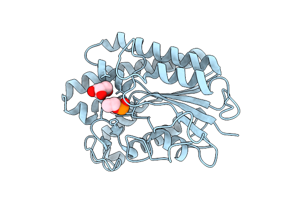
Crystal Structure Of Anae In Apo Form, A Fungal Gdsl-Acetylesterase With Acetylcholinesterase-Like Activity From Aspergillus Niger
Organism: Aspergillus niger
Method: X-RAY DIFFRACTION Release Date: 2025-10-29 Classification: HYDROLASE |
 |
Organism: Aspergillus niger
Method: X-RAY DIFFRACTION Release Date: 2025-10-29 Classification: HYDROLASE Ligands: MES |
 |
Organism: Aspergillus niger
Method: X-RAY DIFFRACTION Release Date: 2025-10-29 Classification: HYDROLASE Ligands: ACT, GOL |
 |
Organism: Aspergillus niger
Method: X-RAY DIFFRACTION Release Date: 2025-10-29 Classification: HYDROLASE |
 |
Organism: Aspergillus niger
Method: X-RAY DIFFRACTION Release Date: 2025-10-29 Classification: HYDROLASE |
 |
Organism: Aspergillus niger
Method: X-RAY DIFFRACTION Release Date: 2025-10-29 Classification: HYDROLASE Ligands: ACT, GOL |
 |
Organism: Aspergillus niger
Method: X-RAY DIFFRACTION Release Date: 2025-10-29 Classification: HYDROLASE Ligands: GOL, DPF |
 |
Organism: Aspergillus niger
Method: X-RAY DIFFRACTION Release Date: 2025-10-29 Classification: HYDROLASE Ligands: MES |
 |
Organism: Aspergillus niger
Method: X-RAY DIFFRACTION Release Date: 2025-10-29 Classification: HYDROLASE Ligands: ACT |
 |
Organism: Aspergillus niger
Method: X-RAY DIFFRACTION Release Date: 2025-10-29 Classification: HYDROLASE Ligands: PPI |
 |
Organism: Aspergillus niger
Method: X-RAY DIFFRACTION Release Date: 2025-10-29 Classification: HYDROLASE Ligands: SO4, MES, DIO |
 |
Organism: Aspergillus niger
Method: X-RAY DIFFRACTION Release Date: 2025-10-29 Classification: HYDROLASE |
 |
Organism: Aspergillus niger
Method: X-RAY DIFFRACTION Release Date: 2025-10-29 Classification: HYDROLASE |
 |
Organism: Aspergillus niger
Method: X-RAY DIFFRACTION Release Date: 2025-10-29 Classification: HYDROLASE |
 |
Organism: Aspergillus niger
Method: X-RAY DIFFRACTION Release Date: 2025-10-29 Classification: HYDROLASE |