Planned Maintenance: Some services may turn out to be unavailable from 15th January, 2026 to 16th January, 2026. We apologize for the inconvenience!

Planned Maintenance: Some services may turn out to be unavailable from 15th January, 2026 to 16th January, 2026. We apologize for the inconvenience!
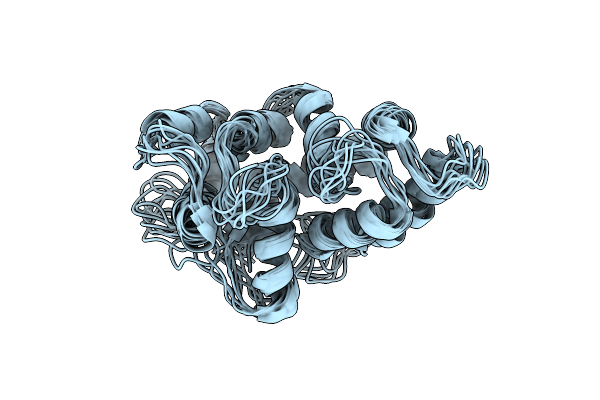 |
Organism: Danio rerio
Method: SOLUTION NMR Release Date: 2024-05-08 Classification: METAL BINDING PROTEIN |
 |
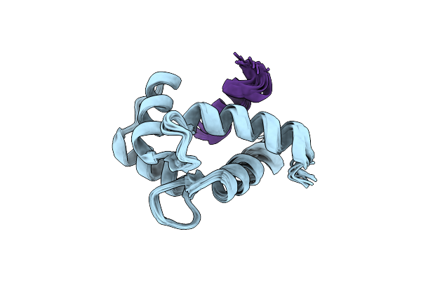
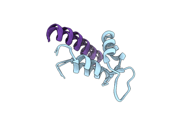
Nmr Structure Of Calmodulin Bound To C-Terminal Site In The Beta-Subunit Of Cyclic Nucleotide-Gated Channel
Organism: Homo sapiens
Method: SOLUTION NMR Release Date: 2022-12-14 Classification: METAL BINDING PROTEIN |
 |
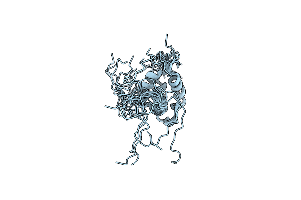
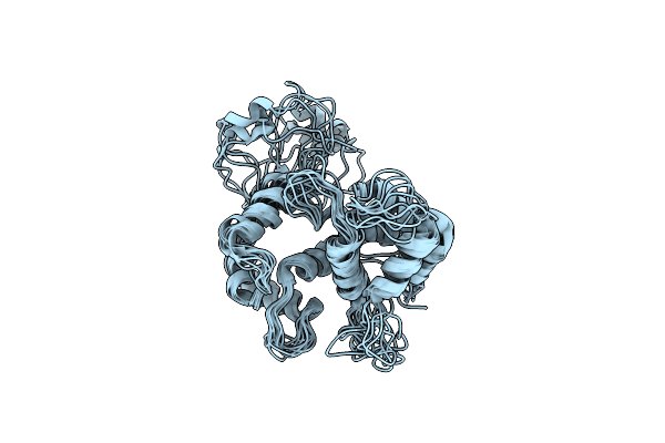
Nmr Structure Of Calmodulin Bound To N-Terminal Site In The Beta-Subunit Of Cyclic Nucleotide-Gated Channel
Organism: Homo sapiens
Method: SOLUTION NMR Release Date: 2022-12-14 Classification: METAL BINDING PROTEIN |
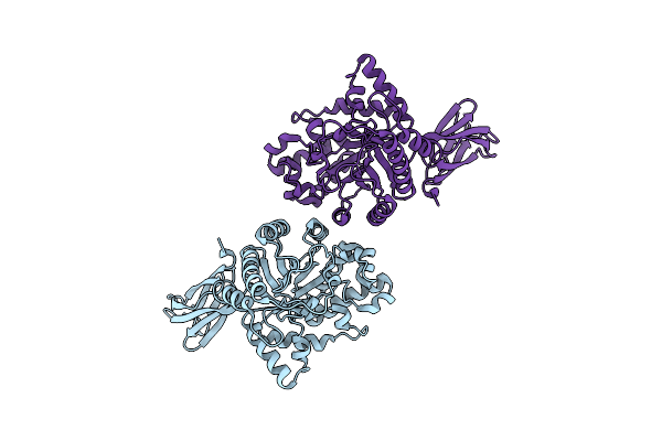 |
Acetivibrio Clariflavus Beta-1,4-Xylanase Of Glycoside Hydrolase Family 10 (Acxyn10A).
Organism: Acetivibrio clariflavus
Method: X-RAY DIFFRACTION Resolution:2.24 Å Release Date: 2022-10-19 Classification: HYDROLASE |
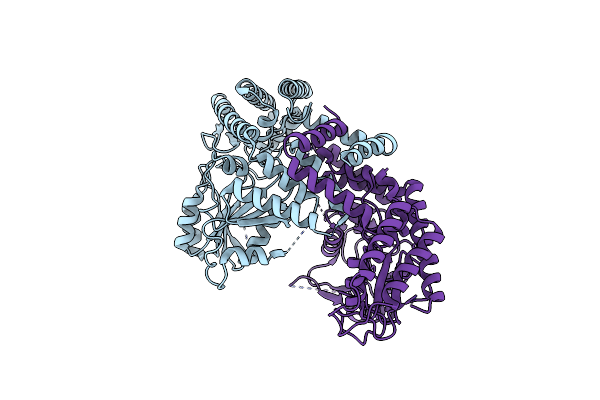 |
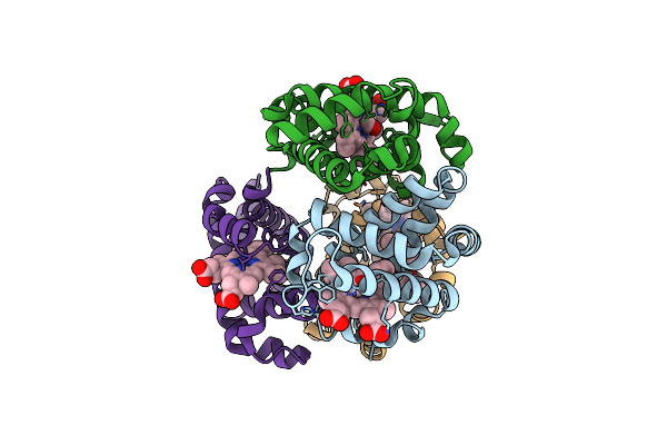
Apo-Structure Of Serine Hydroxymethyltransferase (Pbzb) Involved In Benzobactin Biosynthesis In P. Chlororaphis Subsp. Piscium Dsm 21509
Organism: Pseudomonas chlororaphis subsp. piscium
Method: X-RAY DIFFRACTION Resolution:2.81 Å Release Date: 2022-10-12 Classification: BIOSYNTHETIC PROTEIN |
 |
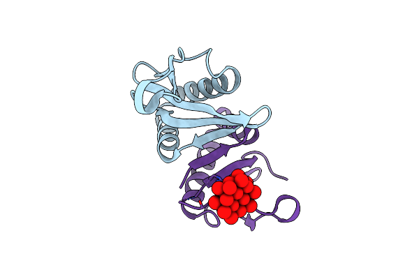
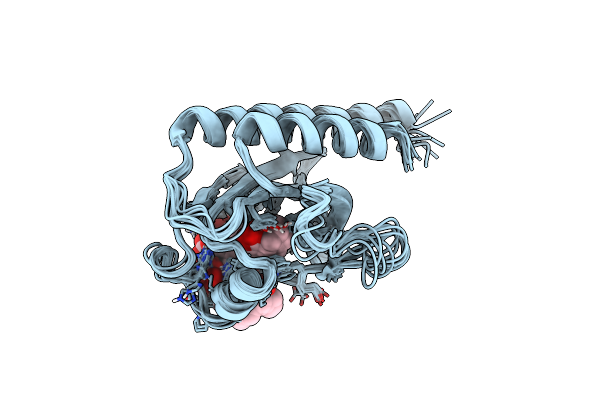
Nmr Structure Of Half-Calcified Calmodulin Mutant (Camef12) Bound To The Iq-Motif Of Cav1.2
Organism: Homo sapiens
Method: SOLUTION NMR Release Date: 2022-07-06 Classification: METAL BINDING PROTEIN Ligands: CA |
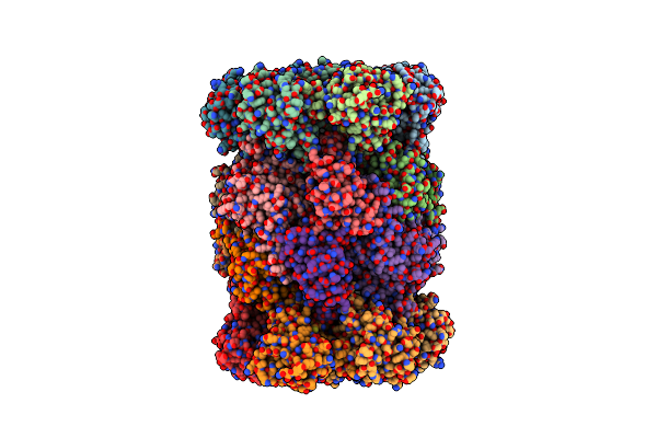 |
Yeast 20S Proteasome In Complex With The Covalently Bound Inhibitor B-Lactone (2R,3S)-3-Isopropyl-4-Oxo-2-Oxetane-Carboxylate (Ioc)
Organism: Saccharomyces cerevisiae
Method: X-RAY DIFFRACTION Release Date: 2022-04-13 Classification: HYDROLASE Ligands: MG, CL, V08 |
 |
Organism: Danio rerio
Method: SOLUTION NMR Release Date: 2021-10-20 Classification: METAL BINDING PROTEIN Ligands: MG |
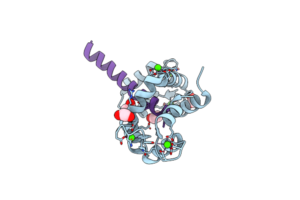 |
Structure Of Calmodulin Bound To The Cardiac Ryanodine Receptor (Ryr2) At Residues: Phe4246 To Val4271
Organism: Homo sapiens
Method: X-RAY DIFFRACTION Resolution:1.65 Å Release Date: 2021-04-07 Classification: METAL BINDING PROTEIN Ligands: CA, EDO |
 |
 |
Organism: Agrobacterium tumefaciens
Method: X-RAY DIFFRACTION Resolution:1.76 Å Release Date: 2019-11-06 Classification: TOXIN Ligands: TEW |
 |
Organism: Xenopus laevis, Oryctolagus cuniculus
Method: SOLUTION NMR Release Date: 2019-09-25 Classification: PROTEIN BINDING |
 |
Organism: Homo sapiens
Method: SOLUTION NMR Release Date: 2019-02-06 Classification: SIGNALING PROTEIN |
 |
Organism: Mus musculus, Homo sapiens
Method: SOLUTION NMR Release Date: 2019-01-16 Classification: SIGNALING PROTEIN |
 |
Organism: Nostoc punctiforme (strain atcc 29133 / pcc 73102)
Method: SOLUTION NMR Release Date: 2018-04-18 Classification: SIGNALING PROTEIN Ligands: CYC |
 |
Organism: Nostoc punctiforme pcc 73102
Method: SOLUTION NMR Release Date: 2018-04-18 Classification: SIGNALING PROTEIN Ligands: CYC |
 |
Organism: Xenopus laevis, Homo sapiens
Method: SOLUTION NMR Release Date: 2017-10-25 Classification: METAL BINDING PROTEIN Ligands: CA |
 |
Organism: Homo sapiens
Method: SOLUTION NMR Release Date: 2017-07-12 Classification: MEMBRANE PROTEIN |
 |
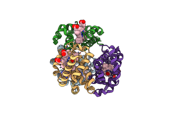
Crystal Structure Of Carbonmonoxy Hemoglobin S (Liganded Sickle Cell Hemoglobin) Complexed With Gbt Compound 31
Organism: Homo sapiens
Method: X-RAY DIFFRACTION Resolution:1.95 Å Release Date: 2017-02-22 Classification: OXYGEN TRANSPORT Ligands: HEM, CMO, 7SJ |
 |
Crystal Structure Of Carbonmonoxy Hemoglobin S (Liganded Sickle Cell Hemoglobin) Complexed With Gbt Compound 6
Organism: Homo sapiens
Method: X-RAY DIFFRACTION Resolution:2.05 Å Release Date: 2017-02-22 Classification: OXYGEN TRANSPORT Ligands: HEM, CMO, 86M |