
Search Count: 222
 |
Organism: Mycobacterium tuberculosis, Synthetic construct
Method: ELECTRON MICROSCOPY Release Date: 2025-10-08 Classification: DNA BINDING PROTEIN Ligands: MG, JHN |
 |
Structure Of Mycobacterium Tuberculosis Steb (Rv1698), A Cell Division Regulator
Organism: Mycobacterium tuberculosis h37rv
Method: X-RAY DIFFRACTION Release Date: 2025-07-30 Classification: CELL CYCLE Ligands: CD, ACY, PG4 |
 |
Organism: Mycobacterium tuberculosis h37rv
Method: X-RAY DIFFRACTION Release Date: 2025-07-30 Classification: CELL CYCLE Ligands: HEZ |
 |
Structure Of Mycobacterium Tuberculosis Stea (Rv1697), A Cell Division Regulator
Organism: Mycobacterium tuberculosis h37rv
Method: X-RAY DIFFRACTION Release Date: 2025-07-30 Classification: CELL CYCLE |
 |
Organism: Corynebacterium glutamicum atcc 13032
Method: X-RAY DIFFRACTION Release Date: 2025-07-30 Classification: CELL CYCLE |
 |
Organism: Mycobacterium tuberculosis, Synthetic construct
Method: ELECTRON MICROSCOPY Release Date: 2025-03-19 Classification: DNA BINDING PROTEIN Ligands: A1H5Q |
 |
Organism: Plasmodium vivax, Synthetic construct
Method: X-RAY DIFFRACTION Resolution:1.60 Å Release Date: 2025-02-05 Classification: CELL INVASION Ligands: CA, NAG, SO4 |
 |
Organism: Plasmodium vivax, Synthetic construct
Method: X-RAY DIFFRACTION Resolution:1.50 Å Release Date: 2024-03-20 Classification: HYDROLASE Ligands: CA, NAG, SO4 |
 |
Organism: Plasmodium vivax, Synthetic construct
Method: X-RAY DIFFRACTION Resolution:1.54 Å Release Date: 2024-03-20 Classification: HYDROLASE Ligands: NAG, CA, SO4 |
 |
Organism: Plasmodium vivax, Synthetic construct
Method: X-RAY DIFFRACTION Resolution:1.77 Å Release Date: 2024-03-20 Classification: HYDROLASE Ligands: CA, NAG, SO4 |
 |
Organism: Plasmodium falciparum
Method: X-RAY DIFFRACTION Resolution:3.09 Å Release Date: 2024-03-06 Classification: HYDROLASE Ligands: CA, PO4 |
 |
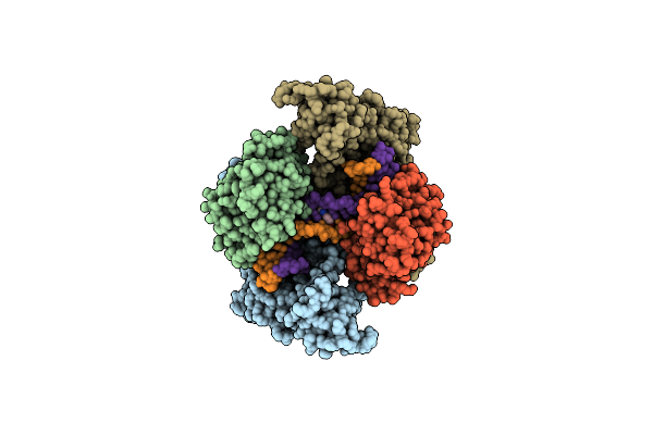
Crystal Structure Of Full-Length, Homohexameric 2-Oxoglutarate Dehydrogenase Kgd From Mycobacterium Smegmatis In Complex With Gara
Organism: Mycolicibacterium smegmatis mc2 155
Method: X-RAY DIFFRACTION Resolution:4.56 Å Release Date: 2023-08-16 Classification: OXIDOREDUCTASE Ligands: MG, CA, TPP |
 |
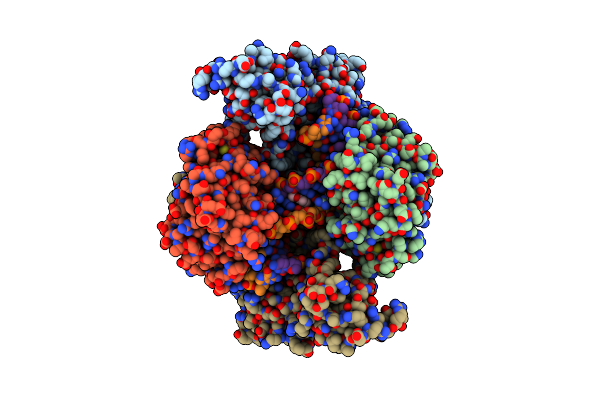
Crystal Structure Of The Homohexameric 2-Oxoglutarate Dehydrogenase Odha From Corynebacterium Glutamicum
Organism: Corynebacterium glutamicum atcc 13032
Method: X-RAY DIFFRACTION Resolution:2.46 Å Release Date: 2023-08-16 Classification: OXIDOREDUCTASE Ligands: EPE, COA, ACO, TPP, MG |
 |
Single Particle Cryo-Em Structure Of The Homohexameric 2-Oxoglutarate Dehydrogenase Odha From Corynebacterium Glutamicum
Organism: Corynebacterium glutamicum atcc 13032
Method: ELECTRON MICROSCOPY Resolution:2.17 Å Release Date: 2023-08-16 Classification: OXIDOREDUCTASE Ligands: MG, ACO, TPP |
 |
Single Particle Cryo-Em Structure Of Homohexameric 2-Oxoglutarate Dehydrogenase Odha From Corynebacterium Glutamicum With Coenzyme A Bound To The E2O Domain
Organism: Corynebacterium glutamicum atcc 13032
Method: ELECTRON MICROSCOPY Resolution:2.17 Å Release Date: 2023-08-16 Classification: OXIDOREDUCTASE Ligands: MG, COA, ACO, TPP |
 |
Single Particle Cryo-Em Structure Of Homohexameric 2-Oxoglutarate Dehydrogenase Odha From Corynebacterium Glutamicum In Complex With The Product Succinyl-Coa
Organism: Corynebacterium glutamicum atcc 13032
Method: ELECTRON MICROSCOPY Release Date: 2023-08-16 Classification: OXIDOREDUCTASE Ligands: MG, ACO, TPP, SCA |
 |
Single Particle Cryo-Em Structure Of Homohexameric 2-Oxoglutarate Dehydrogenase Odha From Corynebacterium Glutamicum Following Reaction With The 2-Oxoglutarate Analogue Succinyl Phosphonate
Organism: Corynebacterium glutamicum atcc 13032
Method: ELECTRON MICROSCOPY Resolution:2.26 Å Release Date: 2023-08-16 Classification: OXIDOREDUCTASE Ligands: MG, ACO, QSP |
 |
Single Particle Cryo-Em Structure Of The Complex Between Corynebacterium Glutamicum Homohexameric 2-Oxoglutarate Dehydrogenase Odha And The Fha-Protein Inhibitor Odhi
Organism: Corynebacterium glutamicum atcc 13032
Method: ELECTRON MICROSCOPY Resolution:2.29 Å Release Date: 2023-08-16 Classification: OXIDOREDUCTASE Ligands: TPP, MG |
 |
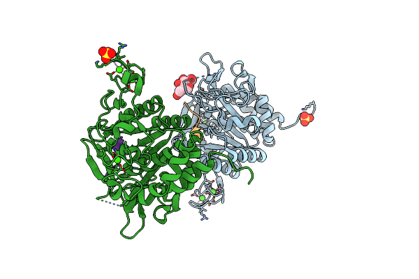
Structure Of The Catalytic Domain Of P. Vivax Sub1 (Triclinic Crystal Form) In Complex With Inhibitor
Organism: Plasmodium vivax, Synthetic construct
Method: X-RAY DIFFRACTION Resolution:1.51 Å Release Date: 2023-07-19 Classification: HYDROLASE Ligands: NAG, CA, SO4 |
 |
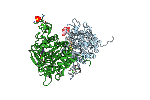
Structure Of The Catalytic Domain Of P. Vivax Sub1 (Triclinic Crystal Form)
Organism: Plasmodium vivax
Method: X-RAY DIFFRACTION Resolution:1.44 Å Release Date: 2023-07-19 Classification: HYDROLASE Ligands: CA, NAG, SO4 |