
Search Count: 63
 |
Organism: Mycosarcoma maydis
Method: X-RAY DIFFRACTION Release Date: 2025-08-20 Classification: UNKNOWN FUNCTION Ligands: SO4 |
 |
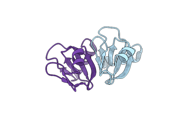
Crystal Structure Of The Tin2-Fold Effector Protein Tue1 From Thecaphora Thlaspeos
Organism: Thecaphora thlaspeos
Method: X-RAY DIFFRACTION Release Date: 2025-06-25 Classification: NUCLEAR PROTEIN |
 |
Organism: Phytophthora infestans
Method: X-RAY DIFFRACTION Resolution:2.30 Å Release Date: 2025-01-15 Classification: HYDROLASE |
 |
Organism: Ustilago maydis 521
Method: X-RAY DIFFRACTION Resolution:1.81 Å Release Date: 2025-01-15 Classification: HYDROLASE |
 |
Organism: Ustilago maydis
Method: X-RAY DIFFRACTION Resolution:1.74 Å Release Date: 2024-10-30 Classification: RNA |
 |
Organism: Ustilago maydis
Method: X-RAY DIFFRACTION Resolution:2.40 Å Release Date: 2024-10-30 Classification: RNA |
 |
Organism: Ustilago maydis
Method: X-RAY DIFFRACTION Resolution:2.00 Å Release Date: 2024-10-30 Classification: RNA BINDING PROTEIN |
 |
Crystal Structure Of The Cerato-Platanin-Like Protein Cpl1 From Ustilago Maydis
Organism: Ustilago maydis 521
Method: X-RAY DIFFRACTION Resolution:1.90 Å Release Date: 2023-05-17 Classification: SUGAR BINDING PROTEIN |
 |
Organism: Ustilago hordei
Method: X-RAY DIFFRACTION Resolution:1.35 Å Release Date: 2023-05-17 Classification: SUGAR BINDING PROTEIN |
 |
Crystal Structure Of The Disulfide Reductase Mera From Staphylococcus Aureus
Organism: Staphylococcus aureus
Method: X-RAY DIFFRACTION Resolution:2.40 Å Release Date: 2023-03-01 Classification: OXIDOREDUCTASE Ligands: FAD, HIS |
 |
Crystal Structure Of A C43S Variant From The Disulfide Reductase Mera From Staphylococcus Aureus
Organism: Staphylococcus aureus
Method: X-RAY DIFFRACTION Resolution:1.60 Å Release Date: 2023-03-01 Classification: OXIDOREDUCTASE Ligands: GOL, FAD, NI |
 |
Organism: Chaetomium thermophilum
Method: X-RAY DIFFRACTION Resolution:2.40 Å Release Date: 2022-09-21 Classification: PROTEIN BINDING Ligands: CIT |
 |
Organism: Methanococcus maripaludis x1
Method: X-RAY DIFFRACTION Resolution:2.30 Å Release Date: 2022-04-27 Classification: METAL BINDING PROTEIN Ligands: MG, BEF |
 |
Organism: Pyrococcus horikoshii (strain atcc 700860 / dsm 12428 / jcm 9974 / nbrc 100139 / ot-3)
Method: X-RAY DIFFRACTION Resolution:2.90 Å Release Date: 2022-04-27 Classification: METAL BINDING PROTEIN |
 |
Crystal Structure Of A Parb E93A Mutant From Myxococcus Xanthus Bound To Cdp And Monothiophosphate
Organism: Myxococcus xanthus (strain dk1622)
Method: X-RAY DIFFRACTION Resolution:1.89 Å Release Date: 2021-09-15 Classification: HYDROLASE Ligands: CDP, TS6, MG, PEG, GOL |
 |
Crystal Structure Of Parb From Myxococcus Xanthus Bound To Cdp And Monothiophosphate
Organism: Myxococcus xanthus (strain dk 1622)
Method: X-RAY DIFFRACTION Resolution:1.90 Å Release Date: 2021-09-08 Classification: DNA BINDING PROTEIN Ligands: GOL, PEG, TS6, CDP, MG |
 |
Crystal Structure Of A Parb Q52A Mutant From Myxococcus Xanthus Bound To Ctpys
Organism: Myxococcus xanthus (strain dk 1622)
Method: X-RAY DIFFRACTION Resolution:1.70 Å Release Date: 2021-09-08 Classification: DNA BINDING PROTEIN Ligands: UFQ, GOL, MG |
 |
Crystal Structure Of The Dna-Binding Protein Rema From Geobacillus Thermodenitrificans
Organism: Geobacillus thermodenitrificans ng80-2
Method: X-RAY DIFFRACTION Resolution:2.29 Å Release Date: 2021-08-25 Classification: DNA BINDING PROTEIN Ligands: SO4 |
 |
Crystal Structure Of A R18W Mutant Of The Dna-Binding Protein Rema From Geobacillus Thermodenitrificans
Organism: Geobacillus thermodenitrificans (strain ng80-2)
Method: X-RAY DIFFRACTION Resolution:2.60 Å Release Date: 2021-08-25 Classification: DNA BINDING PROTEIN |
 |
Crystal Structure Of A R51 R53 Double Mutant Of The Dna-Binding Protein Rema From Geobacillus Thermodenitrificans
Organism: Geobacillus thermodenitrificans (strain ng80-2)
Method: X-RAY DIFFRACTION Resolution:1.80 Å Release Date: 2021-08-25 Classification: DNA BINDING PROTEIN |