
Search Count: 11
 |
Lgl2 Bound To The Apkciota-Par6B Complex In Nucleotide-Free Form. Head Sub-Complex Region Subtracted
Organism: Homo sapiens, Mus musculus
Method: ELECTRON MICROSCOPY Release Date: 2025-07-02 Classification: LIPID BINDING PROTEIN |
 |
Lgl2 Bound To The Apkciota-Par6B Complex In Nucleotide-Free Form. Conformation With Visible Head Sub-Complex.
Organism: Homo sapiens, Mus musculus
Method: ELECTRON MICROSCOPY Release Date: 2025-07-02 Classification: LIPID BINDING PROTEIN |
 |
Organism: Homo sapiens, Mus musculus
Method: ELECTRON MICROSCOPY Release Date: 2025-07-02 Classification: LIPID BINDING PROTEIN Ligands: ADP, MG |
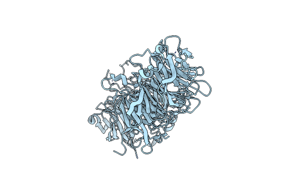 |
Crystal Structure Of The Human Cell Polarity Protein Lethal Giant Larvae 2 (Lgl2). Unphosphorylated, Crystal Form 1.
Organism: Homo sapiens
Method: X-RAY DIFFRACTION Resolution:3.19 Å Release Date: 2019-05-08 Classification: LIPID BINDING PROTEIN Ligands: CL |
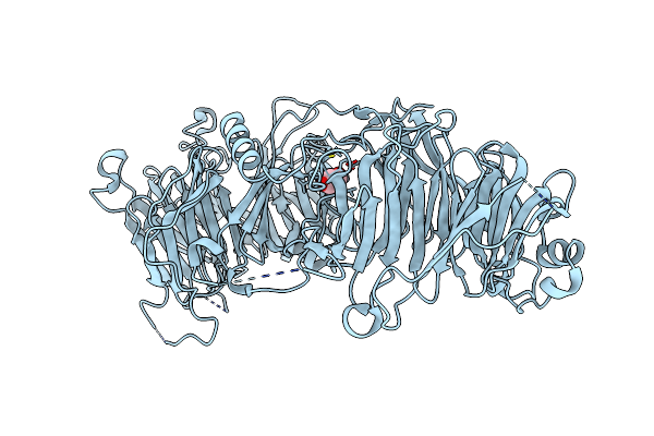 |
Crystal Structure Of The Human Cell Polarity Protein Lethal Giant Larvae 2 (Lgl2). Unphosphorylated, Crystal Form 2.
Organism: Homo sapiens
Method: X-RAY DIFFRACTION Resolution:2.20 Å Release Date: 2019-05-08 Classification: LIPID BINDING PROTEIN Ligands: CL, GOL |
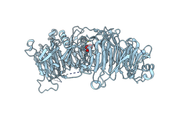 |
Crystal Structure Of The Human Cell Polarity Protein Lethal Giant Larvae 2 (Lgl2). Apkc Phosphorylated, Crystal Form 2.
Organism: Homo sapiens
Method: X-RAY DIFFRACTION Resolution:1.91 Å Release Date: 2019-05-08 Classification: LIPID BINDING PROTEIN Ligands: CL, GOL |
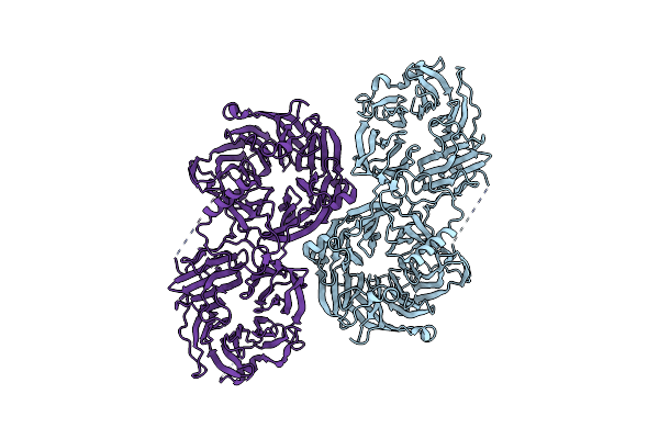 |
Crystal Structure Of The Human Cell Polarity Protein Lethal Giant Larvae 2 (Lgl2). Apkc Phosphorylated, Crystal Form 3.
Organism: Homo sapiens
Method: X-RAY DIFFRACTION Resolution:3.90 Å Release Date: 2019-05-08 Classification: LIPID BINDING PROTEIN |
 |
Organism: Rattus norvegicus
Method: X-RAY DIFFRACTION Resolution:2.00 Å Release Date: 2013-09-11 Classification: METAL BINDING PROTEIN Ligands: SO4, BME, CA, FLC |
 |
Organism: Rattus norvegicus
Method: X-RAY DIFFRACTION Resolution:1.26 Å Release Date: 2013-09-11 Classification: METAL BINDING PROTEIN Ligands: CA, FLC |
 |
Crystal Structure Of The Voltage Dependent Calcium Channel Beta-2 Subunit In Complex With The Cav2.2 I-Ii Linker.
Organism: Oryctolagus cuniculus, Rattus norvegicus
Method: X-RAY DIFFRACTION Resolution:2.00 Å Release Date: 2012-06-13 Classification: TRANSPORT PROTEIN |
 |
Crystal Structure Of The Voltage Dependent Calcium Channel Beta-2 Subunit In Complex With The Cav1.2 I-Ii Linker.
Organism: Oryctolagus cuniculus
Method: X-RAY DIFFRACTION Resolution:1.95 Å Release Date: 2012-06-13 Classification: TRANSPORT PROTEIN Ligands: BR |