
Search Count: 49
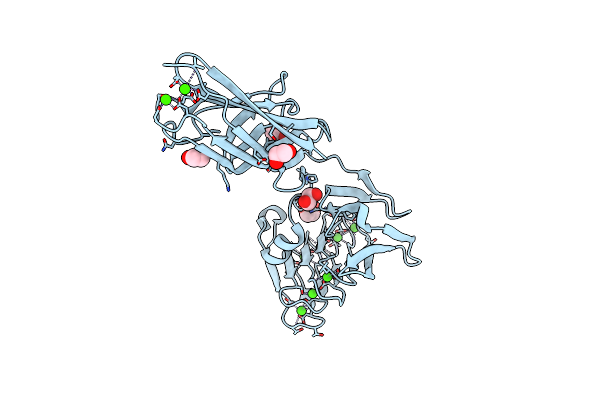 |
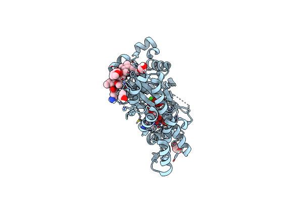
Crystal Structure Of Repeats-In-Toxin-Like Domain From Aeromonas Hydrophila
Organism: Aeromonas hydrophila
Method: X-RAY DIFFRACTION Resolution:1.95 Å Release Date: 2025-05-14 Classification: CELL ADHESION Ligands: EDO, CA |
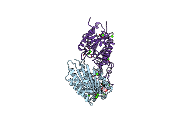 |
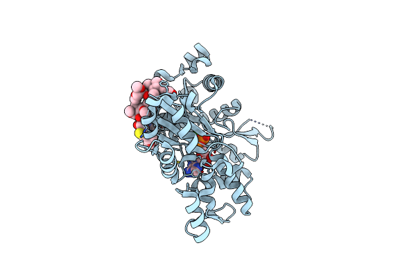
Crystal Structure Of Vwfa Domain From Large Adhesion Protein Of Aeromonas Hydrophila
Organism: Aeromonas hydrophila
Method: X-RAY DIFFRACTION Resolution:1.40 Å Release Date: 2025-05-14 Classification: CELL ADHESION Ligands: CA, GOL |
 |
Organism: Candida albicans
Method: X-RAY DIFFRACTION Release Date: 2024-09-04 Classification: MOTOR PROTEIN |
 |
Organism: Candida albicans, Sus scrofa
Method: ELECTRON MICROSCOPY Release Date: 2022-07-20 Classification: MOTOR PROTEIN Ligands: GTP, MG, GDP, TA1, ANP |
 |
Organism: Candida albicans, Sus scrofa
Method: ELECTRON MICROSCOPY Release Date: 2022-07-20 Classification: MOTOR PROTEIN Ligands: AF3, ADP, MG, GTP, GDP, TA1 |
 |
Organism: Candida albicans, Sus scrofa
Method: ELECTRON MICROSCOPY Release Date: 2022-07-20 Classification: MOTOR PROTEIN Ligands: GTP, MG, GDP, TA1, ANP |
 |
Organism: Candida albicans, Homo sapiens, Sus scrofa
Method: ELECTRON MICROSCOPY Release Date: 2022-07-20 Classification: MOTOR PROTEIN Ligands: MG, ANP, GTP, GDP, TA1 |
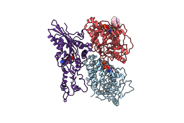 |
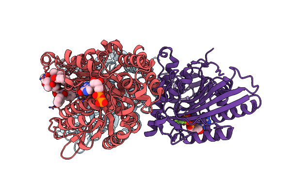
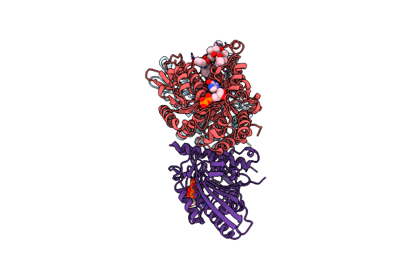
Cakip3[2-482] - Amp-Pnp In Complex With A Dolastatin-10-Stabilized Tubulin Ring
Organism: Candida albicans, Sus scrofa
Method: ELECTRON MICROSCOPY Release Date: 2022-07-20 Classification: MOTOR PROTEIN Ligands: GTP, MG, GDP, SR6, ANP |
 |
Organism: Candida albicans, Sus scrofa
Method: ELECTRON MICROSCOPY Release Date: 2022-07-13 Classification: MOTOR PROTEIN Ligands: GTP, MG, GDP, TA1 |
 |
Organism: Candida albicans, Homo sapiens, Sus scrofa
Method: ELECTRON MICROSCOPY Release Date: 2022-07-13 Classification: MOTOR PROTEIN Ligands: GTP, MG, GDP, TA1 |
 |
Organism: Candida albicans (strain sc5314 / atcc mya-2876)
Method: X-RAY DIFFRACTION Resolution:2.01 Å Release Date: 2022-02-09 Classification: MOTOR PROTEIN Ligands: ADP, MG |
 |
Organism: Oryctolagus cuniculus
Method: X-RAY DIFFRACTION Resolution:1.70 Å Release Date: 2020-10-21 Classification: CONTRACTILE PROTEIN Ligands: CA, ATP, LAB, EDO, TFJ |
 |
Organism: Oryctolagus cuniculus
Method: X-RAY DIFFRACTION Resolution:2.20 Å Release Date: 2018-11-21 Classification: STRUCTURAL PROTEIN Ligands: JQV, CA, ADP |
 |
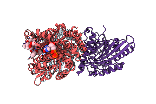
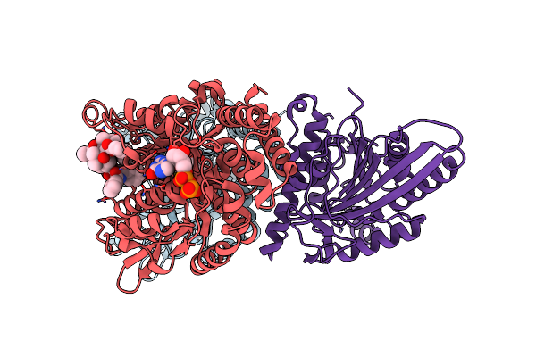
Crystal Structure Of A Curved Tubulin Complex Induced By The Kinesin-13 Kif2A
Organism: Escherichia coli, Homo sapiens, Bos taurus
Method: X-RAY DIFFRACTION Resolution:3.51 Å Release Date: 2018-05-23 Classification: MOTOR PROTEIN Ligands: GTP, MG, GDP, PO4, ANP |
 |
Organism: Pseudomonas brassicacearum
Method: X-RAY DIFFRACTION Resolution:1.90 Å Release Date: 2018-03-07 Classification: OXIDOREDUCTASE Ligands: FE |
 |
Organism: Marinomonas primoryensis
Method: X-RAY DIFFRACTION Resolution:2.00 Å Release Date: 2017-09-06 Classification: CELL ADHESION Ligands: CA |
 |
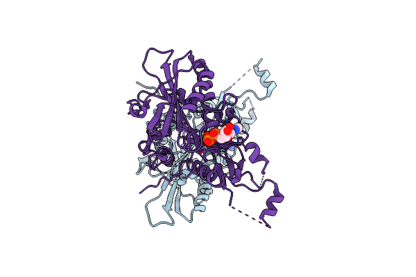
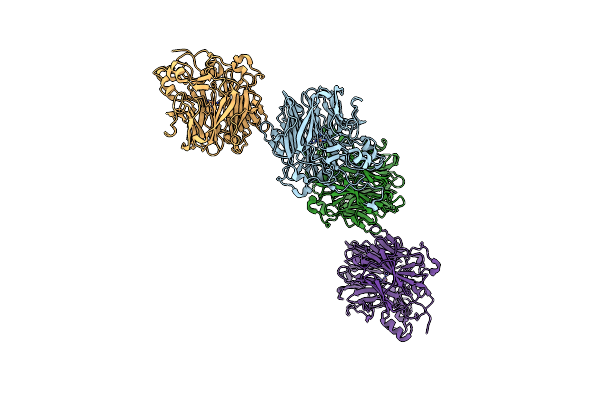
Structural Insight Into Host Cell Surface Retention Of A 1.5-Mda Bacterial Ice-Binding Adhesin
Organism: Marinomonas primoryensis
Method: X-RAY DIFFRACTION Resolution:2.00 Å Release Date: 2017-07-19 Classification: CELL ADHESION Ligands: CA, MG, EDO |
 |
Organism: Marinomonas primoryensis
Method: X-RAY DIFFRACTION Resolution:1.35 Å Release Date: 2017-07-19 Classification: CELL ADHESION Ligands: CA |
 |
Organism: Marinomonas primoryensis
Method: SOLUTION NMR Release Date: 2017-06-28 Classification: Antifreeze protein, Cell adhesion |
 |
Organism: Marinomonas primoryensis
Method: X-RAY DIFFRACTION Resolution:1.03 Å Release Date: 2017-06-07 Classification: CELL ADHESION Ligands: CA, GLC, BGC |