
Search Count: 105
 |
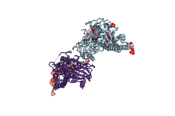
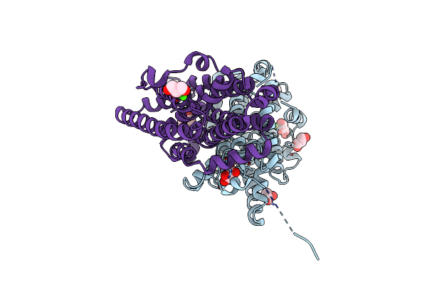
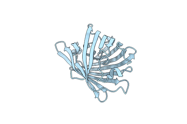
Serial Femtosecond X-Ray Structure Of A Fluorescence Optimized Bathy Phytochrome Pairfp2 Derived From Wild-Type Agp2 In Its Pfr State (I0A).
Organism: Agrobacterium fabrum str. c58
Method: X-RAY DIFFRACTION Resolution:2.15 Å Release Date: 2025-05-14 Classification: SIGNALING PROTEIN Ligands: EL5, SO4, CL, EDO |
 |
Serial Femtosecond X-Ray Structure Of A Fluorescence Optimized Bathy Phytochrome Pairfp2 Derived From Wild-Type Agp2 In Its Pfr State (I0B).
Organism: Agrobacterium fabrum str. c58
Method: X-RAY DIFFRACTION Resolution:2.20 Å Release Date: 2025-05-14 Classification: SIGNALING PROTEIN Ligands: EL5, SO4 |
 |
Serial Femtosecond X-Ray Structure Of A Fluorescence Optimized Bathy Phytochrome Pairfp2 Derived From Wild-Type Agp2 In I1 Intermediate State.
Organism: Agrobacterium fabrum str. c58
Method: X-RAY DIFFRACTION Resolution:2.54 Å Release Date: 2025-05-14 Classification: SIGNALING PROTEIN Ligands: EL5, SO4 |
 |
Serial Femtosecond X-Ray Structure Of A Fluorescence Optimized Bathy Phytochrome Pairfp2 Derived From Wild-Type Agp2 In I2 Intermediate State.
Organism: Agrobacterium fabrum str. c58
Method: X-RAY DIFFRACTION Resolution:2.43 Å Release Date: 2025-05-14 Classification: SIGNALING PROTEIN Ligands: EL5, SO4, PGE, PEG, CL |
 |
Serial Femtosecond X-Ray Structure Of A Fluorescence Optimized Bathy Phytochrome Pairfp2 Derived From Wild-Type Agp2 In I3 Intermediate State.
Organism: Agrobacterium fabrum str. c58
Method: X-RAY DIFFRACTION Resolution:2.40 Å Release Date: 2025-05-14 Classification: SIGNALING PROTEIN Ligands: EL5, SO4, GOL, PEG |
 |
Serial Femtosecond X-Ray Structure Of A Fluorescence Optimized Bathy Phytochrome Pairfp2 Derived From Wild-Type Agp2 In I4 Intermediate State.
Organism: Agrobacterium fabrum str. c58
Method: X-RAY DIFFRACTION Resolution:2.30 Å Release Date: 2025-05-14 Classification: SIGNALING PROTEIN Ligands: EL5, SO4, CL, PEG |
 |
Serial Femtosecond X-Ray Structure Of A Fluorescence Optimized Bathy Phytochrome Pairfp2 Derived From Wild-Type Agp2 In I5 Intermediate State.
Organism: Agrobacterium fabrum str. c58
Method: X-RAY DIFFRACTION Resolution:2.43 Å Release Date: 2025-05-14 Classification: SIGNALING PROTEIN Ligands: EL5, SO4, CL |
 |
Serial Femtosecond X-Ray Structure Of A Fluorescence Optimized Bathy Phytochrome Pairfp2 Derived From Wild-Type Agp2 In I6 Intermediate State.
Organism: Agrobacterium fabrum str. c58
Method: X-RAY DIFFRACTION Resolution:2.49 Å Release Date: 2025-05-14 Classification: SIGNALING PROTEIN Ligands: EL5, SO4, CL |
 |
Serial Femtosecond X-Ray Structure Of A Fluorescence Optimized Bathy Phytochrome Pairfp2 Derived From Wild-Type Agp2 In I7 Intermediate State.
Organism: Agrobacterium fabrum str. c58
Method: X-RAY DIFFRACTION Resolution:2.80 Å Release Date: 2025-05-14 Classification: SIGNALING PROTEIN Ligands: EL5, SO4, PEG |
 |
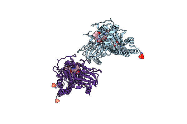
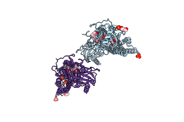
Crystal Structure Of Actinonin-Bound Pdf1 And The Computationally Designed Dbact553_1 Protein Binder
Organism: Pseudomonas aeruginosa, Synthetic construct
Method: X-RAY DIFFRACTION Resolution:1.88 Å Release Date: 2024-10-30 Classification: DE NOVO PROTEIN Ligands: ZN, BB2, FMT, PO4, K |
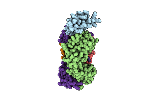 |
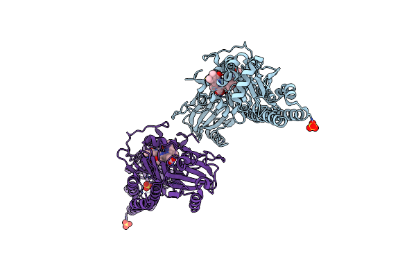
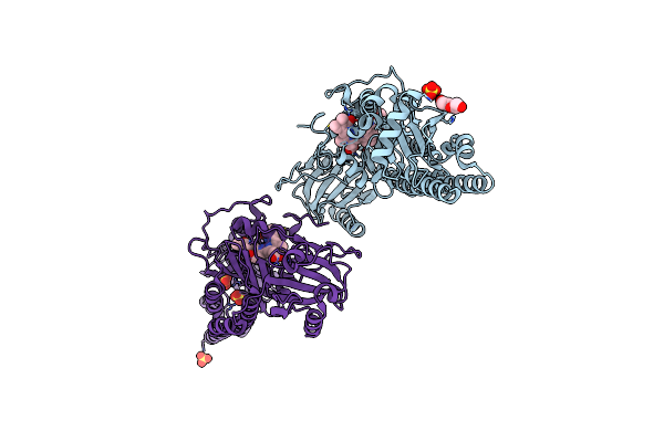
Progesterone-Bound Db3 Fab In Complex With Computationally Designed Dbpro1156_2 Protein Binder
Organism: Synthetic construct
Method: ELECTRON MICROSCOPY Release Date: 2024-10-30 Classification: DE NOVO PROTEIN Ligands: STR |
 |
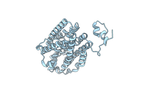
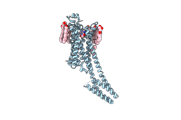
Organism: Tursiops truncatus
Method: ELECTRON MICROSCOPY Release Date: 2023-12-13 Classification: MEMBRANE PROTEIN Ligands: CL |
 |
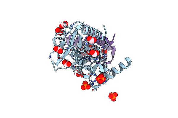
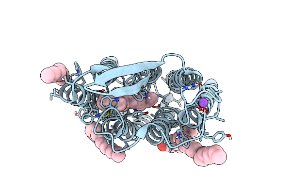
Time-Resolved Sfx-Xfel Crystal Structure Of Cyp121 Bound With Cyy Reacted With Peracetic Acid For 200 Milliseconds
Organism: Mycobacterium tuberculosis h37rv
Method: X-RAY DIFFRACTION Resolution:1.85 Å Release Date: 2023-11-22 Classification: OXIDOREDUCTASE Ligands: SO4, HEM, YTT, PEO |
 |
Organism: Mycobacterium tuberculosis h37rv
Method: X-RAY DIFFRACTION Resolution:1.65 Å Release Date: 2023-11-22 Classification: OXIDOREDUCTASE Ligands: SO4, HEM, YTT |
 |
Ribonucleotide Reductase Class Ie R2 From Mesoplasma Florum, Catalytically Active Radical State Solved By Xfel
Organism: Mesoplasma florum l1
Method: X-RAY DIFFRACTION Resolution:1.50 Å Release Date: 2023-11-01 Classification: OXIDOREDUCTASE |
 |
Ribonucleotide Reductase Class Ie R2 From Mesoplasma Florum, Radical-Lost Ground State
Organism: Mesoplasma florum l1
Method: X-RAY DIFFRACTION Resolution:1.35 Å Release Date: 2023-11-01 Classification: OXIDOREDUCTASE Ligands: CA, GOL |
 |
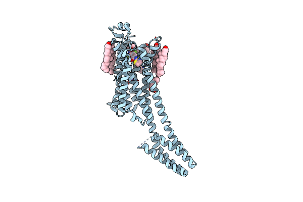
Room-Temperature Structure Of The Stabilised A2A-Theophylline Complex Determined By Synchrotron Serial Crystallography
Organism: Homo sapiens
Method: X-RAY DIFFRACTION Resolution:3.45 Å Release Date: 2023-08-30 Classification: MEMBRANE PROTEIN Ligands: TEP, CLR, OLA, OLC, NA |
 |
Room-Temperature Structure Of The Stabilised A2A-Luaa47070 Complex Determined By Synchrotron Serial Crystallography
Organism: Homo sapiens
Method: X-RAY DIFFRACTION Resolution:3.50 Å Release Date: 2023-08-30 Classification: MEMBRANE PROTEIN Ligands: 9Y2, CLR, OLA, NA |
 |
Room Temperature Structure Of Archaerhodopsin-3 Obtained 110 Ns After Photoexcitation
Organism: Halorubrum sodomense
Method: X-RAY DIFFRACTION Resolution:1.80 Å Release Date: 2023-06-14 Classification: PROTON TRANSPORT Ligands: RET, DD9, R16, PLM, CA, NA, MG, CL |
 |
Organism: Lobophyllia hemprichii
Method: X-RAY DIFFRACTION Resolution:1.75 Å Release Date: 2023-01-11 Classification: FLUORESCENT PROTEIN |