
Search Count: 17
 |
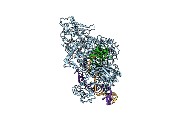
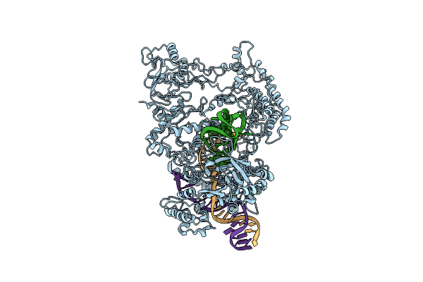
Organism: Gallus gallus, Synthetic construct
Method: ELECTRON MICROSCOPY Release Date: 2024-07-31 Classification: DNA BINDING PROTEIN |
 |
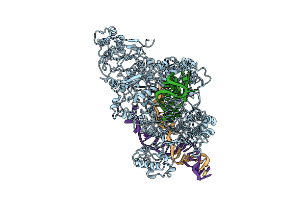
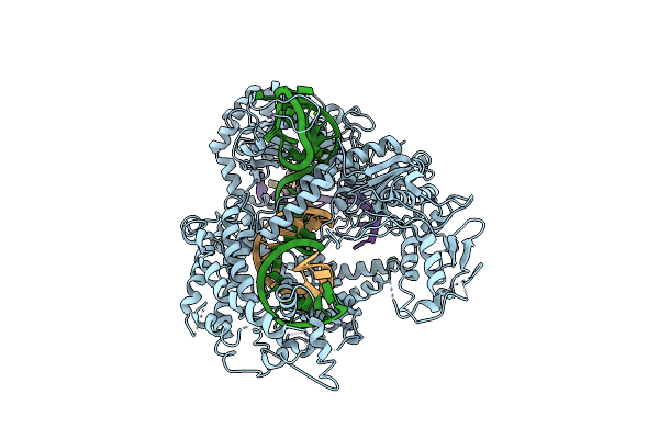
Organism: Gallus gallus, Synthetic construct
Method: ELECTRON MICROSCOPY Release Date: 2024-07-31 Classification: DNA BINDING PROTEIN |
 |
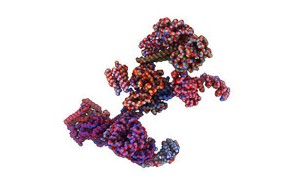
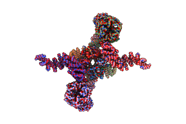
Structure Of The Dna-Bound Fancd2-Fanci Complex Containing Phosphomimetic Fanci
Organism: Gallus gallus, Synthetic construct
Method: ELECTRON MICROSCOPY Release Date: 2022-09-07 Classification: DNA BINDING PROTEIN |
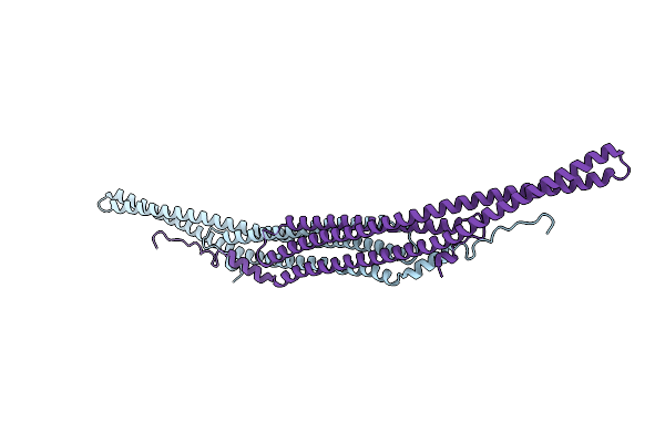 |
Organism: Homo sapiens
Method: X-RAY DIFFRACTION Resolution:1.97 Å Release Date: 2022-02-23 Classification: SIGNALING PROTEIN |
 |
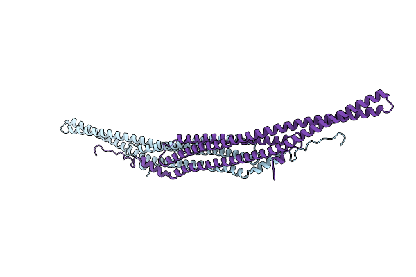
Crystal Structure Of The F-Bar Domain Of Pstipip1 Bound To The Cth Domain Of The Phosphatase Lyp
Organism: Homo sapiens
Method: X-RAY DIFFRACTION Resolution:2.15 Å Release Date: 2022-02-23 Classification: SIGNALING PROTEIN Ligands: GOL |
 |
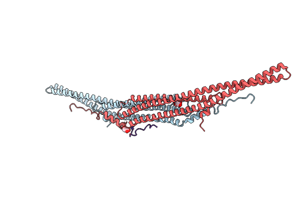
Organism: Homo sapiens
Method: X-RAY DIFFRACTION Resolution:2.14 Å Release Date: 2022-02-23 Classification: SIGNALING PROTEIN |
 |
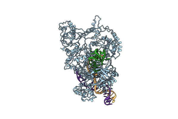
Organism: Gallus gallus, Homo sapiens, Synthetic construct
Method: ELECTRON MICROSCOPY Release Date: 2020-02-19 Classification: DNA BINDING PROTEIN |
 |
Organism: Gallus gallus
Method: ELECTRON MICROSCOPY Release Date: 2020-02-19 Classification: DNA BINDING PROTEIN |
 |
Organism: Gallus gallus
Method: ELECTRON MICROSCOPY Release Date: 2020-02-19 Classification: DNA BINDING PROTEIN |
 |
 |
 |
Organism: Francisella tularensis subsp. novicida u112
Method: ELECTRON MICROSCOPY Release Date: 2018-12-19 Classification: HYDROLASE |
 |
Organism: Francisella tularensis subsp. novicida (strain u112), Francisella tularensis subsp. novicida u112
Method: ELECTRON MICROSCOPY Release Date: 2018-12-19 Classification: HYDROLASE |
 |
Organism: Francisella tularensis subsp. novicida (strain u112), Francisella tularensis subsp. novicida u112
Method: ELECTRON MICROSCOPY Release Date: 2018-12-19 Classification: HYDROLASE |
 |
Organism: Francisella tularensis subsp. novicida u112
Method: ELECTRON MICROSCOPY Release Date: 2018-12-19 Classification: HYDROLASE |
 |
Organism: Francisella tularensis subsp. novicida u112
Method: ELECTRON MICROSCOPY Release Date: 2018-12-19 Classification: HYDROLASE Ligands: MG |
 |
Organism: Francisella tularensis subsp. novicida u112
Method: X-RAY DIFFRACTION Resolution:3.00 Å Release Date: 2017-06-21 Classification: HYDROLASE Ligands: MG |