
Search Count: 34
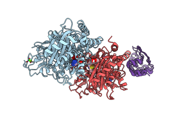 |
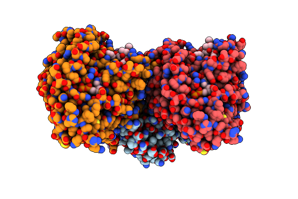
Organism: Synthetic construct, Bos taurus
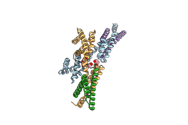
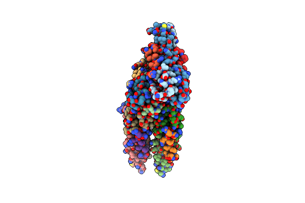
Method: X-RAY DIFFRACTION Resolution:1.82 Å Release Date: 2022-03-30 Classification: CELL CYCLE Ligands: GTP, MG, CA, GDP, I8R |
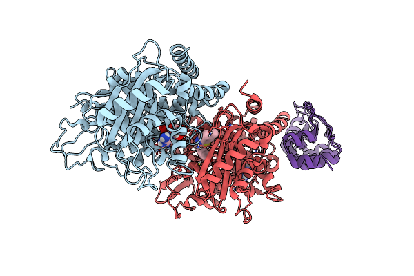 |
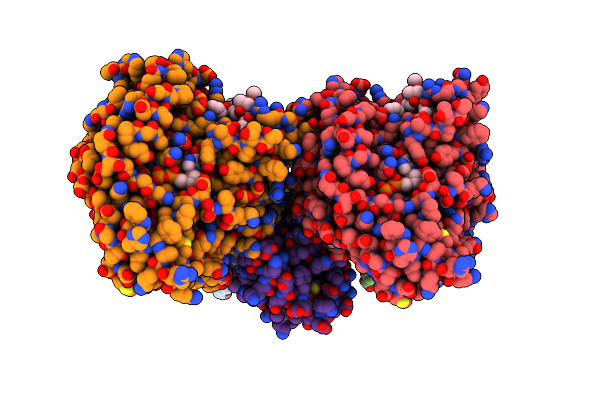
Organism: Synthetic construct, Bos taurus
Method: X-RAY DIFFRACTION Resolution:2.36 Å Release Date: 2022-03-30 Classification: CELL CYCLE Ligands: GTP, MG, GDP, I8N |
 |
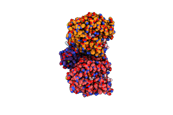
Organism: Synthetic construct, Bos taurus
Method: X-RAY DIFFRACTION Resolution:1.75 Å Release Date: 2020-12-23 Classification: CELL CYCLE Ligands: GTP, MG, GDP, QRN |
 |
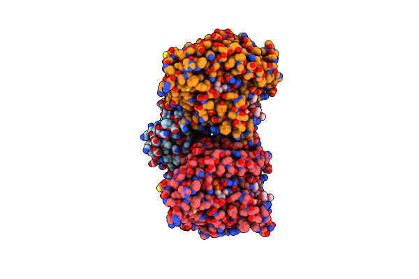
Organism: Synthetic construct, Bos taurus
Method: X-RAY DIFFRACTION Resolution:2.04 Å Release Date: 2020-12-23 Classification: CELL CYCLE Ligands: GTP, MG, GDP, QRQ |
 |
Organism: Homo sapiens
Method: X-RAY DIFFRACTION Resolution:2.19 Å Release Date: 2020-05-27 Classification: CELL ADHESION Ligands: EDO |
 |
Organism: Homo sapiens
Method: X-RAY DIFFRACTION Resolution:2.30 Å Release Date: 2020-05-27 Classification: CELL ADHESION |
 |
Organism: Homo sapiens
Method: ELECTRON MICROSCOPY Release Date: 2019-11-27 Classification: STRUCTURAL PROTEIN Ligands: GTP, MG, GDP, TA1 |
 |
Organism: Naegleria gruberi, Homo sapiens
Method: ELECTRON MICROSCOPY Release Date: 2019-11-27 Classification: STRUCTURAL PROTEIN Ligands: GTP, MG, GDP, TA1 |
 |
Organism: Naegleria gruberi, Homo sapiens
Method: ELECTRON MICROSCOPY Release Date: 2019-11-27 Classification: STRUCTURAL PROTEIN Ligands: GTP, MG, GDP, TA1 |
 |
Organism: Homo sapiens
Method: ELECTRON MICROSCOPY Release Date: 2019-11-27 Classification: STRUCTURAL PROTEIN Ligands: GTP, MG, GDP, TA1 |
 |
Organism: Mus musculus
Method: X-RAY DIFFRACTION Resolution:2.00 Å Release Date: 2019-08-07 Classification: HYDROLASE Ligands: PEG, EDO |
 |
Organism: Mus musculus
Method: X-RAY DIFFRACTION Resolution:1.70 Å Release Date: 2018-07-11 Classification: HYDROLASE Ligands: EDO, PEG |
 |
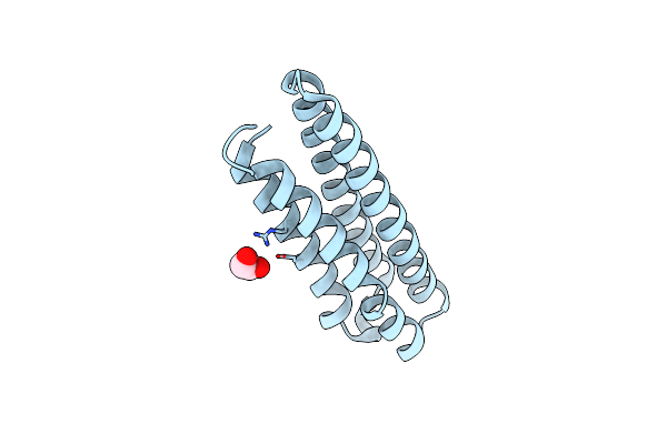
Organism: Homo sapiens
Method: X-RAY DIFFRACTION Resolution:1.20 Å Release Date: 2018-05-30 Classification: STRUCTURAL PROTEIN |
 |
Organism: Rattus norvegicus, Gallus gallus, Bos taurus
Method: X-RAY DIFFRACTION Resolution:2.40 Å Release Date: 2017-10-11 Classification: CELL CYCLE Ligands: GTP, MG, CA, GOL, GDP, 6FS, MES, ACP |
 |
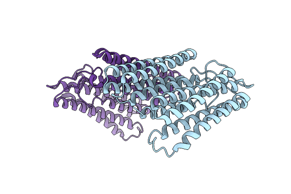
Organism: Mus musculus
Method: X-RAY DIFFRACTION Resolution:1.40 Å Release Date: 2017-10-04 Classification: STRUCTURAL PROTEIN |
 |
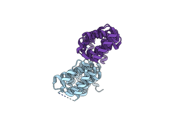
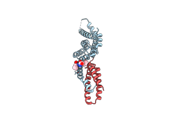
Mechanism Of Microtubule Minus-End Recognition And Protection By Camsap Proteins
Organism: Mus musculus, Bos taurus
Method: ELECTRON MICROSCOPY Release Date: 2017-10-04 Classification: STRUCTURAL PROTEIN Ligands: GTP, MG, GDP, TA1 |
 |
Mechanism Of Microtubule Minus-End Recognition And Protection By Camsap Proteins
Organism: Homo sapiens, Bos taurus
Method: ELECTRON MICROSCOPY Release Date: 2017-10-04 Classification: MOTOR PROTEIN Ligands: GTP, MG, GDP, TA1 |
 |
Mechanism Of Microtubule Minus-End Recognition And Protection By Camsap Proteins
Organism: Homo sapiens, Bos taurus
Method: ELECTRON MICROSCOPY Release Date: 2017-10-04 Classification: TRANSPORT PROTEIN Ligands: GTP, MG, GDP, TA1 |
 |
Organism: Homo sapiens, Saccharomyces cerevisiae
Method: X-RAY DIFFRACTION Resolution:2.30 Å Release Date: 2017-06-14 Classification: microtubule binding protein |
 |
Complex Structure Between P60N/P80C Katanin And A Peptide Derived From Aspm
Organism: Mus musculus
Method: X-RAY DIFFRACTION Resolution:1.50 Å Release Date: 2017-04-26 Classification: HYDROLASE |