
Search Count: 23
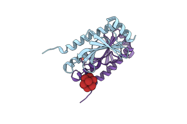 |
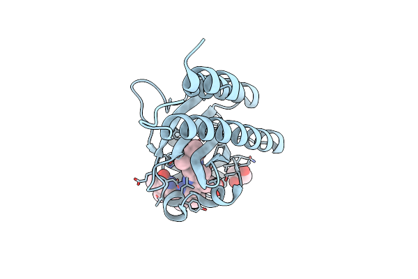
Crystal Structure Of The Type Iii Secretion Chaperone Veca From Vibrio Parahaemolyticus
Organism: Vibrio parahaemolyticus
Method: X-RAY DIFFRACTION Release Date: 2025-06-25 Classification: CHAPERONE Ligands: PO4, TBR |
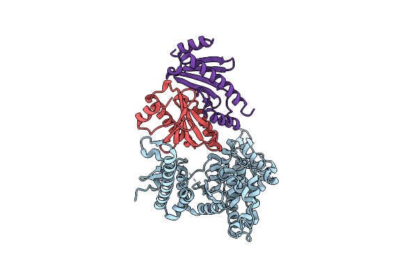 |
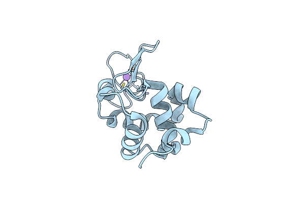
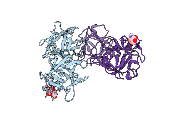
Crystal Structure Of The Virulence Effector Vepa In Complex With Its Secretion Chaperone Veca
Organism: Vibrio parahaemolyticus
Method: X-RAY DIFFRACTION Release Date: 2025-06-25 Classification: TOXIN |
 |
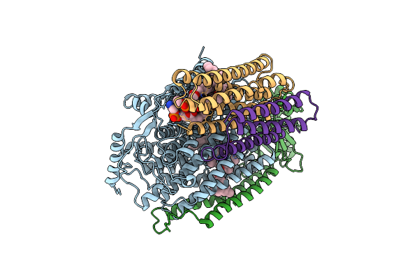
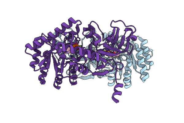
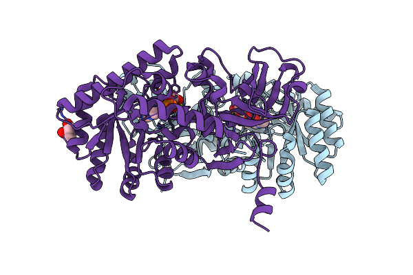
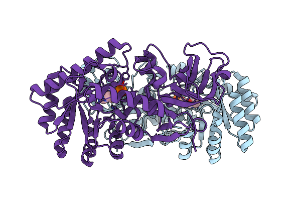
Crystal Structure Of Bovine Heart Cytochrome C Oxidase, Apo Structure With Dmso
Organism: Bos taurus
Method: X-RAY DIFFRACTION Resolution:2.20 Å Release Date: 2022-12-21 Classification: OXIDOREDUCTASE Ligands: HEA, CU, MG, NA, PER, PGV, TGL, CUA, CDL, CHD, PEK, PSC, ZN, DMU |
 |
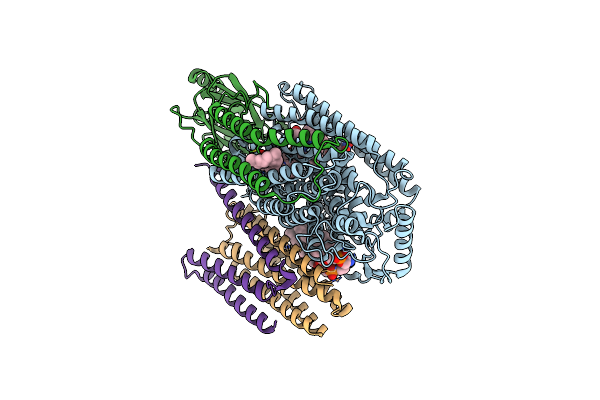
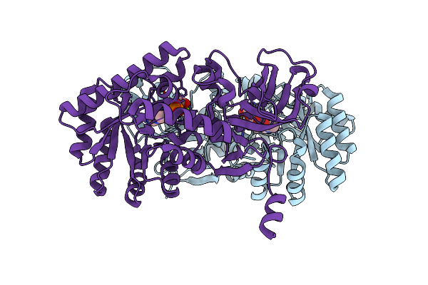
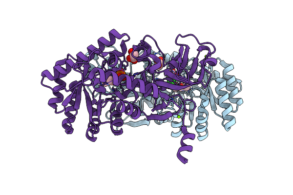
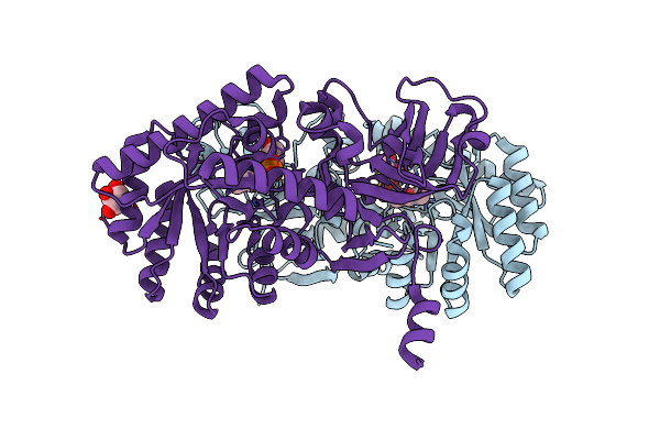
Crystal Structure Of Bovine Heart Cytochrome C Oxidase, The Structure Complexed With An Allosteric Inhibitor T113
Organism: Bos taurus
Method: X-RAY DIFFRACTION Resolution:2.20 Å Release Date: 2022-12-21 Classification: OXIDOREDUCTASE Ligands: HEA, CU, MG, NA, PER, TGL, J6X, CUA, CHD, PSC, PEK, PGV, CDL, DMU, ZN |
 |
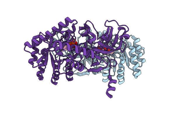
Cryo-Em Structure Of Cytochrome Bo3 From Escherichia Coli, Apo Structure With Dmso
Organism: Escherichia coli
Method: ELECTRON MICROSCOPY Release Date: 2022-12-21 Classification: OXIDOREDUCTASE Ligands: HEO, HEM, CU, PEE, UNX |
 |
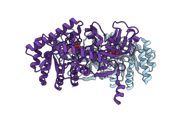
Cryo-Em Structure Of Cytochrome Bo3 From Escherichia Coli, The Structure Complexed With An Allosteric Inhibitor N4
Organism: Escherichia coli
Method: ELECTRON MICROSCOPY Release Date: 2022-12-21 Classification: OXIDOREDUCTASE Ligands: HEO, HEM, CU, PEE, JYR, UNX |
 |
Organism: Mus musculus
Method: ELECTRON MICROSCOPY Release Date: 2022-01-19 Classification: DNA BINDING PROTEIN |
 |
Organism: Mus musculus
Method: ELECTRON MICROSCOPY Release Date: 2022-01-19 Classification: DNA BINDING PROTEIN |
 |
Crystal Structure Of A Far-Red Light-Absorbing Form Of Anpixjg2_Bv4 In Complex With Biliverdin
Organism: Nostoc sp.
Method: X-RAY DIFFRACTION Resolution:1.60 Å Release Date: 2019-04-17 Classification: SIGNALING PROTEIN Ligands: BLA, GOL |
 |
Xfel Structure Of Hen Egg-White Lysozyme Solved Using A Droplet Injector At Sacla
Organism: Gallus gallus
Method: X-RAY DIFFRACTION Resolution:2.30 Å Release Date: 2016-04-13 Classification: HYDROLASE Ligands: CL, NA |
 |
Organism: Delftia sp.
Method: X-RAY DIFFRACTION Resolution:1.70 Å Release Date: 2015-03-11 Classification: LYASE Ligands: PLP, MG |
 |
D-Threo-3-Hydroxyaspartate Dehydratase H351A Mutant Complexed With 2-Amino Maleic Acid
Organism: Delftia sp.
Method: X-RAY DIFFRACTION Resolution:1.80 Å Release Date: 2015-03-11 Classification: LYASE Ligands: PLP, MG, 2KZ |
 |
D-Threo-3-Hydroxyaspartate Dehydratase H351A Mutant Complexed With L-Erythro-3-Hydroxyaspartate
Organism: Delftia sp.
Method: X-RAY DIFFRACTION Resolution:1.90 Å Release Date: 2015-03-11 Classification: LYASE Ligands: PLP, MG, BH2, GOL |
 |
Crystal Structure Of A Potent Anti-Hiv Lectin Actinohivin In Complex With Alpha-1,2-Mannotriose
Organism: Actinomycete sp.
Method: X-RAY DIFFRACTION Resolution:1.40 Å Release Date: 2015-03-04 Classification: SUGAR BINDING PROTEIN |
 |
Organism: Delftia
Method: X-RAY DIFFRACTION Resolution:1.50 Å Release Date: 2015-01-28 Classification: LYASE Ligands: PLP, MG, GOL, CL |
 |
D-Threo-3-Hydroxyaspartate Dehydratase From Delftia Sp. Ht23 Complexed With D-Erythro-3-Hydroxyaspartate
Organism: Delftia
Method: X-RAY DIFFRACTION Resolution:1.50 Å Release Date: 2015-01-28 Classification: LYASE Ligands: PLP, MG, 999 |
 |
D-Threo-3-Hydroxyaspartate Dehydratase From Delftia Sp. Ht23 Complexed With D-Allothreonine
Organism: Delftia
Method: X-RAY DIFFRACTION Resolution:1.60 Å Release Date: 2015-01-28 Classification: LYASE Ligands: PLP, MG, 2TL |
 |
D-Threo-3-Hydroxyaspartate Dehydratase From Delftia Sp. Ht23 In The Metal-Free Form
Organism: Delftia
Method: X-RAY DIFFRACTION Resolution:2.30 Å Release Date: 2015-01-28 Classification: LYASE Ligands: PLP |
 |
Organism: Delftia
Method: X-RAY DIFFRACTION Resolution:1.55 Å Release Date: 2015-01-28 Classification: LYASE Ligands: PLP, GOL |
 |
Ha1 (Ha33) Subcomponent Of Botulinum Type C Progenitor Toxin Complexed With N-Acetylgalactosamine, Bound At Site Ii
Organism: Clostridium botulinum
Method: X-RAY DIFFRACTION Resolution:1.80 Å Release Date: 2011-06-01 Classification: TOXIN Ligands: NGA |